Department of Neurology, University of Miami Brain Endowment Bank, University of Miami Miller School of Medicine, Miami, Florida, USA.
Evelyn F. McKnight Brain Institute, Department of Neurology, University of Miami Miller School of Medicine, Miami, Florida, USA.
Brain Pathol. 2023 Jul;33(4):e13142. doi: 10.1111/bpa.13142. Epub 2022 Dec 29.
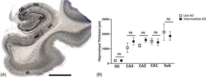
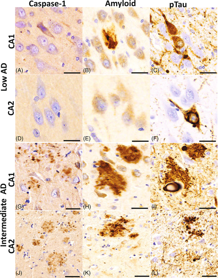
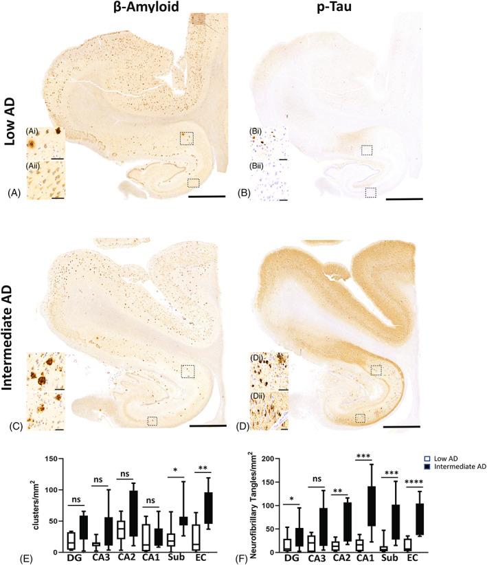
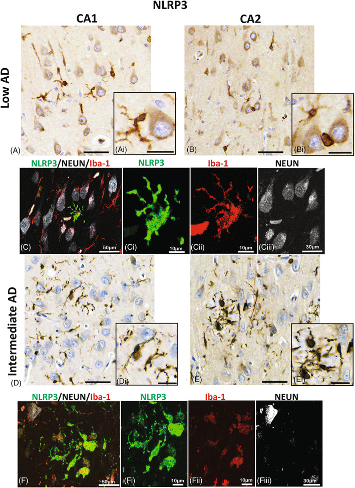
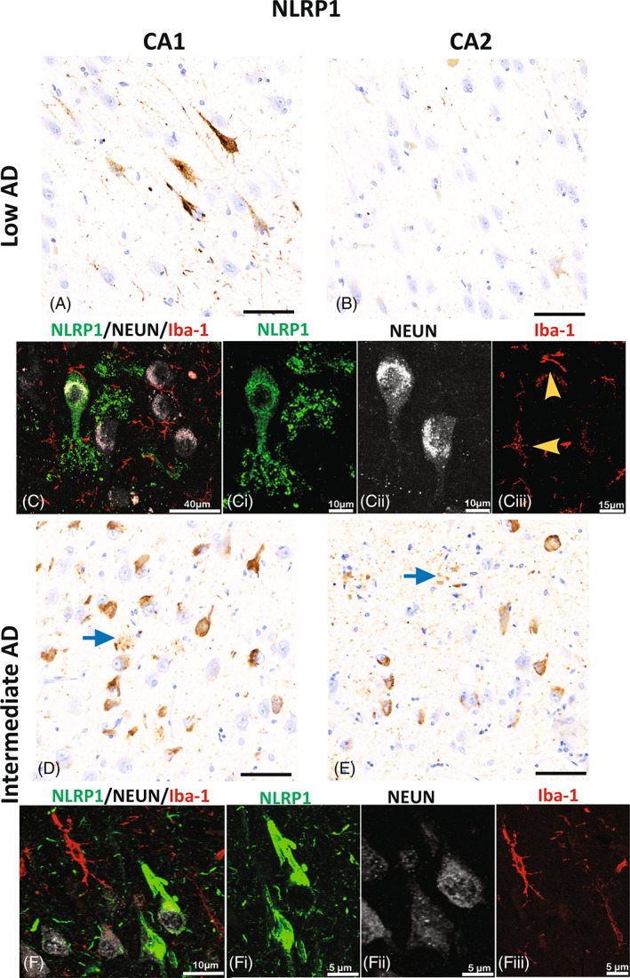
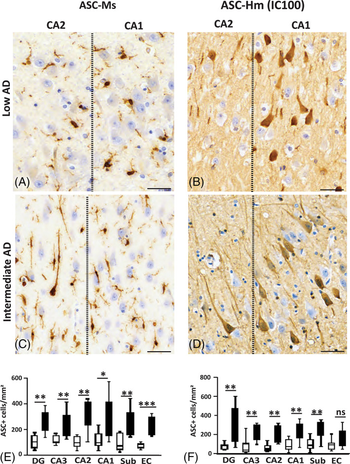
Alzheimer's disease (AD) is a progressive neurodegenerative disease that destroys memory and cognitive function. Inflammasome activation has been suggested to play a critical role in the neuroinflammatory response in AD progression, but the cell-type expression of inflammasome proteins in the brain has not been fully characterized. In this study, we used samples from the hippocampus formation, the subiculum, and the entorhinal cortex brain from 17 donors with low-level AD pathology and 17 intermediate AD donors to assess the expression of inflammasome proteins. We performed analysis of hippocampal thickness, β-amyloid plaques, and hyperphosphorylated tau to ascertain the cellular pathological changes that occur between low and intermediate AD pathology. Next, we determined changes in the cells that express the inflammasome sensor proteins NOD-like receptor proteins (NLRP) 1 and 3, and caspase-1. In addition, we stained section with IC100, a humanized monoclonal antibody directed against the inflammasome adaptor protein apoptosis-associated speck-like protein containing a caspase recruitment domain (ASC), and a commercially available anti-ASC antibody. Our results indicate that hippocampal cortical thickness did not significantly change between low and intermediate AD pathology, but there was an increase in pTau and β-amyloid clusters in intermediate AD cases. NLRP3 was identified mainly in microglial populations, whereas NLRP1 was seen in neuronal cytoplasmic regions. There was a significant increase of ASC in neurons labeled by IC100, whereas microglia in the hippocampus and subiculum were labeled with the commercial anti-ASC antibody. Caspase-1 was present in the parenchyma in the CA regions where amyloid and pTau were identified. Together, our results indicate increased inflammasome protein expression in the early pathological stages of AD, that IC100 identifies neurons in early stages of AD and that ASC expression correlates with Aβ and pTau in postmortem AD brains.
阿尔茨海默病(AD)是一种进行性神经退行性疾病,会破坏记忆和认知功能。炎症小体的激活被认为在 AD 进展的神经炎症反应中起关键作用,但大脑中炎症小体蛋白的细胞类型表达尚未完全表征。在这项研究中,我们使用了来自 17 名低水平 AD 病理供体和 17 名中度 AD 供体的海马结构、下托和内嗅皮层脑组织样本,评估了炎症小体蛋白的表达。我们分析了海马厚度、β-淀粉样斑块和过度磷酸化的 tau,以确定低水平 AD 病理和中度 AD 病理之间发生的细胞病理变化。接下来,我们确定了表达炎症小体传感器蛋白 NOD 样受体蛋白(NLRP)1 和 3 以及半胱氨酸蛋白酶-1 的细胞的变化。此外,我们用 IC100 染色切片,IC100 是一种针对炎症小体衔接蛋白含有半胱氨酸蛋白酶募集结构域(ASC)的凋亡相关斑点样蛋白的人源化单克隆抗体,以及一种市售的抗 ASC 抗体。我们的结果表明,在低水平 AD 病理和中度 AD 病理之间,海马皮质厚度没有显著变化,但在中度 AD 病例中,pTau 和β-淀粉样斑块簇增加。NLRP3 主要在小胶质细胞群体中被识别,而 NLRP1 则在神经元细胞质区域中被识别。在由 IC100 标记的神经元中,ASC 的表达显著增加,而海马和下托中的小胶质细胞则用市售的抗 ASC 抗体标记。Caspase-1 存在于淀粉样蛋白和 pTau 被识别的 CA 区域的实质中。总之,我们的研究结果表明,AD 的早期病理阶段炎症小体蛋白表达增加,IC100 可识别 AD 的早期阶段的神经元,而 ASC 的表达与 AD 死后大脑中的 Aβ 和 pTau 相关。