• 文献检索
  • 文档翻译
  • 深度研究
  • 学术资讯
  • Suppr Zotero 插件Zotero 插件
  • 邀请有礼
  • 套餐&价格
  • 历史记录
应用&插件
Suppr Zotero 插件Zotero 插件浏览器插件Mac 客户端Windows 客户端微信小程序
定价
高级版会员购买积分包购买API积分包
服务
文献检索文档翻译深度研究API 文档MCP 服务
关于我们
关于 Suppr公司介绍联系我们用户协议隐私条款
关注我们

Suppr 超能文献

核心技术专利:CN118964589B侵权必究
粤ICP备2023148730 号-1Suppr @ 2026

文献检索

告别复杂PubMed语法,用中文像聊天一样搜索,搜遍4000万医学文献。AI智能推荐,让科研检索更轻松。

立即免费搜索

文件翻译

保留排版,准确专业,支持PDF/Word/PPT等文件格式,支持 12+语言互译。

免费翻译文档

深度研究

AI帮你快速写综述,25分钟生成高质量综述,智能提取关键信息,辅助科研写作。

立即免费体验

小鼠微型计算机断层扫描(micro-CT)中肺部和肺肿瘤的自动分割

Automated segmentation of lungs and lung tumors in mouse micro-CT scans.

作者信息

Ferl Gregory Z, Barck Kai H, Patil Jasmine, Jemaa Skander, Malamut Evelyn J, Lima Anthony, Long Jason E, Cheng Jason H, Junttila Melissa R, Carano Richard A D

机构信息

Preclinical & Translational PKPD, Genentech, South San Francisco, CA 94080, USA.

Department of Translational Imaging, Genentech, South San Francisco, CA 94080, USA.

出版信息

iScience. 2022 Dec 5;25(12):105712. doi: 10.1016/j.isci.2022.105712. eCollection 2022 Dec 22.

DOI:10.1016/j.isci.2022.105712
PMID:36582483
原文链接:https://pmc.ncbi.nlm.nih.gov/articles/PMC9792881/
Abstract

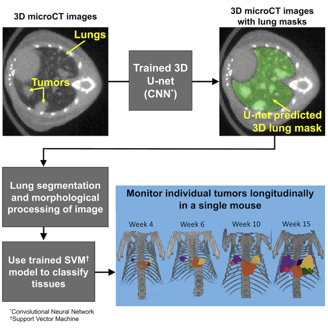
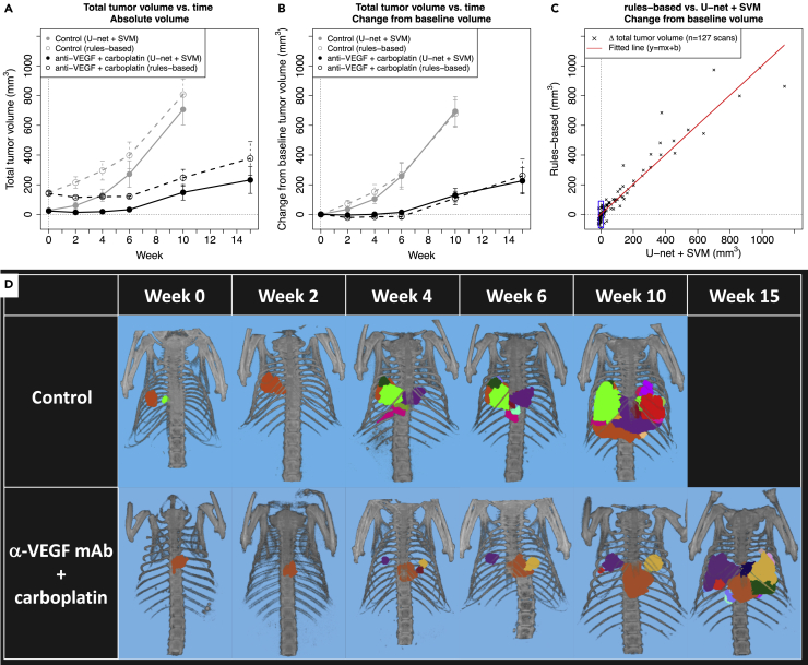
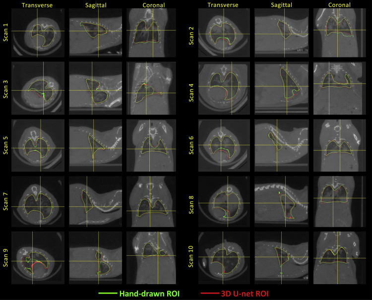
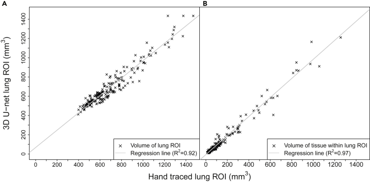
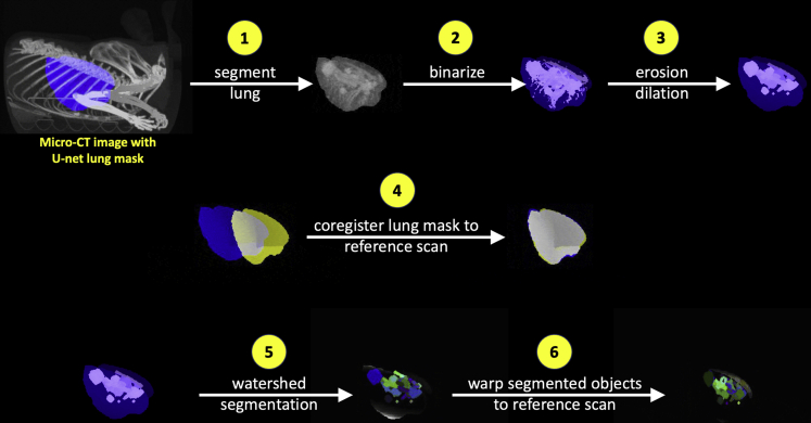
Here, we have developed an automated image processing algorithm for segmenting lungs and individual lung tumors in micro-computed tomography (micro-CT) scans of mouse models of non-small cell lung cancer and lung fibrosis. Over 3000 scans acquired across multiple studies were used to train/validate a 3D U-net lung segmentation model and a Support Vector Machine (SVM) classifier to segment individual lung tumors. The U-net lung segmentation algorithm can be used to estimate changes in soft tissue volume within lungs (primarily tumors and blood vessels), whereas the trained SVM is able to discriminate between tumors and blood vessels and identify individual tumors. The trained segmentation algorithms (1) significantly reduce time required for lung and tumor segmentation, (2) reduce bias and error associated with manual image segmentation, and (3) facilitate identification of individual lung tumors and objective assessment of changes in lung and individual tumor volumes under different experimental conditions.

摘要

在此,我们开发了一种自动图像处理算法,用于在非小细胞肺癌和肺纤维化小鼠模型的微型计算机断层扫描(micro-CT)中分割肺部和单个肺肿瘤。在多项研究中获取的3000多张扫描图像用于训练/验证一个3D U-net肺部分割模型和一个支持向量机(SVM)分类器,以分割单个肺肿瘤。U-net肺部分割算法可用于估计肺内软组织体积的变化(主要是肿瘤和血管),而经过训练的SVM能够区分肿瘤和血管并识别单个肿瘤。经过训练的分割算法(1)显著减少了肺部和肿瘤分割所需的时间,(2)减少了与手动图像分割相关的偏差和误差,(3)便于识别单个肺肿瘤,并客观评估不同实验条件下肺和单个肿瘤体积的变化。

https://cdn.ncbi.nlm.nih.gov/pmc/blobs/9cf1/9792881/d7461fd2d4be/gr4.jpg
https://cdn.ncbi.nlm.nih.gov/pmc/blobs/9cf1/9792881/effcc469705d/fx1.jpg
https://cdn.ncbi.nlm.nih.gov/pmc/blobs/9cf1/9792881/4190e11e7c85/gr1.jpg
https://cdn.ncbi.nlm.nih.gov/pmc/blobs/9cf1/9792881/916de94d6e4c/gr2.jpg
https://cdn.ncbi.nlm.nih.gov/pmc/blobs/9cf1/9792881/09281d510346/gr3.jpg
https://cdn.ncbi.nlm.nih.gov/pmc/blobs/9cf1/9792881/d7461fd2d4be/gr4.jpg

相似文献

1
Automated segmentation of lungs and lung tumors in mouse micro-CT scans.小鼠微型计算机断层扫描(micro-CT)中肺部和肺肿瘤的自动分割
iScience. 2022 Dec 5;25(12):105712. doi: 10.1016/j.isci.2022.105712. eCollection 2022 Dec 22.
2
Applying deep learning to segmentation of murine lung tumors in pre-clinical micro-computed tomography.将深度学习应用于临床前微型计算机断层扫描中鼠肺肿瘤的分割。
Transl Oncol. 2024 Feb;40:101833. doi: 10.1016/j.tranon.2023.101833. Epub 2023 Dec 20.
3
Interactive lung segmentation in abnormal human and animal chest CT scans.异常人类和动物胸部CT扫描中的交互式肺部分割
Med Phys. 2014 Aug;41(8):081915. doi: 10.1118/1.4890597.
4
A computational pipeline for quantification of pulmonary infections in small animal models using serial PET-CT imaging.使用连续 PET-CT 成像对小动物模型中的肺部感染进行定量分析的计算流程。
EJNMMI Res. 2013 Jul 23;3(1):55. doi: 10.1186/2191-219X-3-55.
5
Automatic Organ Segmentation for CT Scans Based on Super-Pixel and Convolutional Neural Networks.基于超像素和卷积神经网络的 CT 扫描自动器官分割。
J Digit Imaging. 2018 Oct;31(5):748-760. doi: 10.1007/s10278-018-0052-4.
6
Segmentation of lung parenchyma in CT images using CNN trained with the clustering algorithm generated dataset.基于聚类算法生成数据集训练的 CNN 对 CT 图像中的肺实质进行分割。
Biomed Eng Online. 2019 Jan 3;18(1):2. doi: 10.1186/s12938-018-0619-9.
7
Three-dimensional lung tumor segmentation from x-ray computed tomography using sparse field active models.基于稀疏域主动模型的 X 射线计算机断层扫描三维肺肿瘤分割。
Med Phys. 2012 Feb;39(2):851-65. doi: 10.1118/1.3676687.
8
Deep Learning Based Automated Orthotopic Lung Tumor Segmentation in Whole-Body Mouse CT-Scans.基于深度学习的全身小鼠CT扫描中原位肺肿瘤自动分割
Cancers (Basel). 2021 Sep 13;13(18):4585. doi: 10.3390/cancers13184585.
9
A fully automatic segmentation algorithm for CT lung images based on random forest.基于随机森林的 CT 肺图像全自动分割算法。
Med Phys. 2020 Feb;47(2):518-529. doi: 10.1002/mp.13939. Epub 2019 Dec 29.
10
Rule and Neural Network-Based Image Segmentation of Mice Vertebrae Images.基于规则和神经网络的小鼠椎骨图像分割
Cureus. 2022 Jul 25;14(7):e27247. doi: 10.7759/cureus.27247. eCollection 2022 Jul.

引用本文的文献

1
Imaging technologies in experimental pulmonary fibrosis research: essential tool for enhanced translational relevance.实验性肺纤维化研究中的成像技术:增强转化相关性的重要工具。
Eur Respir Rev. 2025 Sep 3;34(177). doi: 10.1183/16000617.0012-2025. Print 2025 Jul.
2
Unveiling Another Dimension: Advanced Visualization of Cancer Invasion and Metastasis via Micro-CT Imaging.揭示另一个维度:通过微型计算机断层扫描成像对癌症侵袭和转移进行高级可视化
Cancers (Basel). 2025 Mar 28;17(7):1139. doi: 10.3390/cancers17071139.
3
A Thorough Review of the Clinical Applications of Artificial Intelligence in Lung Cancer.

本文引用的文献

1
Deep Learning-based Automatic Lung Segmentation on Multiresolution CT Scans from Healthy and Fibrotic Lungs in Mice.基于深度学习的小鼠健康肺和纤维化肺多分辨率CT扫描的自动肺分割
Radiol Artif Intell. 2022 Jan 12;4(2):e210095. doi: 10.1148/ryai.210095. eCollection 2022 Mar.
2
Deep learning-based segmentation of the thorax in mouse micro-CT scans.基于深度学习的小鼠 micro-CT 扫描中胸部的分割。
Sci Rep. 2022 Feb 2;12(1):1822. doi: 10.1038/s41598-022-05868-7.
3
Detection of Lung Nodules in Micro-CT Imaging Using Deep Learning.基于深度学习的微 CT 成像中肺结节检测。
人工智能在肺癌临床应用的全面综述
Cancers (Basel). 2025 Mar 4;17(5):882. doi: 10.3390/cancers17050882.
4
Hybrid transformer-CNN and LSTM model for lung disease segmentation and classification.用于肺病分割与分类的混合变压器-CNN和LSTM模型
PeerJ Comput Sci. 2024 Dec 13;10:e2444. doi: 10.7717/peerj-cs.2444. eCollection 2024.
5
Automated recognition and segmentation of lung cancer cytological images based on deep learning.基于深度学习的肺癌细胞学图像自动识别与分割
PLoS One. 2025 Jan 31;20(1):e0317996. doi: 10.1371/journal.pone.0317996. eCollection 2025.
6
The Value of Micro-CT in the Diagnosis of Lung Carcinoma: A Radio-Histopathological Perspective.微计算机断层扫描在肺癌诊断中的价值:放射-组织病理学视角
Diagnostics (Basel). 2023 Oct 20;13(20):3262. doi: 10.3390/diagnostics13203262.
7
Semi-automated micro-computed tomography lung segmentation and analysis in mouse models.小鼠模型中的半自动微型计算机断层扫描肺部分割与分析
MethodsX. 2023 Apr 20;10:102198. doi: 10.1016/j.mex.2023.102198. eCollection 2023.
Tomography. 2021 Aug 7;7(3):358-372. doi: 10.3390/tomography7030032.
4
Mouse lung automated segmentation tool for quantifying lung tumors after micro-computed tomography.用于在微计算机断层扫描后量化肺部肿瘤的小鼠肺部自动分割工具。
PLoS One. 2021 Jun 17;16(6):e0252950. doi: 10.1371/journal.pone.0252950. eCollection 2021.
5
Cancer statistics for the year 2020: An overview.2020年癌症统计数据概述。
Int J Cancer. 2021 Apr 5. doi: 10.1002/ijc.33588.
6
nnU-Net: a self-configuring method for deep learning-based biomedical image segmentation.nnU-Net:一种基于深度学习的生物医学图像分割的自配置方法。
Nat Methods. 2021 Feb;18(2):203-211. doi: 10.1038/s41592-020-01008-z. Epub 2020 Dec 7.
7
Deep learning-enabled multi-organ segmentation in whole-body mouse scans.基于深度学习的全身体积小鼠扫描中多器官分割。
Nat Commun. 2020 Nov 6;11(1):5626. doi: 10.1038/s41467-020-19449-7.
8
Prediction of major torso organs in low-contrast micro-CT images of mice using a two-stage deeply supervised fully convolutional network.使用两阶段深度监督全卷积网络预测小鼠低对比度 micro-CT 图像中的主要躯干器官。
Phys Med Biol. 2019 Dec 19;64(24):245014. doi: 10.1088/1361-6560/ab59a4.
9
Bridging the translational gap: Implementation of multimodal small animal imaging strategies for tumor burden assessment in a co-clinical trial.弥合转化差距:联合临床试验中多模态小动物成像策略在肿瘤负担评估中的应用。
PLoS One. 2019 Apr 8;14(4):e0207555. doi: 10.1371/journal.pone.0207555. eCollection 2019.
10
Muscle specific kinase (MuSK) activation preserves neuromuscular junctions in the diaphragm but is not sufficient to provide a functional benefit in the SOD1 mouse model of ALS.肌肉特异性激酶 (MuSK) 的激活可以保护膈肌中的神经肌肉接头,但不足以在 SOD1 肌萎缩侧索硬化症小鼠模型中提供功能益处。
Neurobiol Dis. 2019 Apr;124:340-352. doi: 10.1016/j.nbd.2018.12.002. Epub 2018 Dec 4.