Dijkstra H Paul, Mc Auliffe Sean, Ardern Clare L, Kemp Joanne L, Mosler Andrea Britt, Price Amy, Blazey Paul, Richards Dawn, Farooq Abdulaziz, Serner Andreas, McNally Eugene, Mascarenhas Vasco, Willy Richard W, Oke Jason L, Khan Karim M, Glyn-Jones Sion, Clarke Mike, Greenhalgh Trisha
Department of Medical Education, Aspetar Orthopaedic and Sports Medicine Hospital, Doha, Qatar
Department for Continuing Education, University of Oxford, Oxford, UK.
Br J Sports Med. 2022 Dec 6;57(6):325-41. doi: 10.1136/bjsports-2022-106085.
Primary cam morphology is a mostly benign bony prominence that develops at the femoral head-neck junction of the hip, but it is highly prevalent in many athlete populations. In the small proportion of athletes for whom it is not benign, the resulting hip osteoarthritis can be debilitating. Clinicians, athletes, patients and researchers do not yet agree on important primary cam morphology elements. We aimed to ascertain and improve the level of agreement on primary cam morphology definitions, terminology, taxonomy and imaging outcome measures.
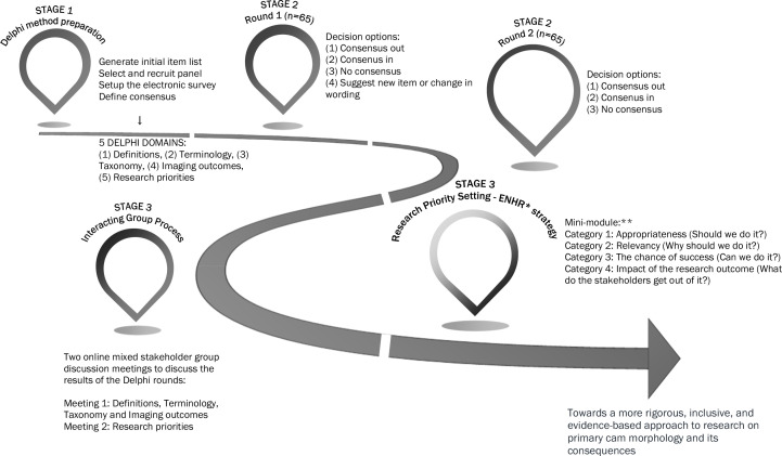
To collect and aggregate informed opinions, an expert panel-the Young Athlete's Hip Research Collaborative-rated primary cam morphology definition, terminology, taxonomy and imaging outcome statements through an online Delphi exercise followed by an online meeting to explore areas of tension and dissent. Reporting followed Conducting and REporting DElphi Studies.
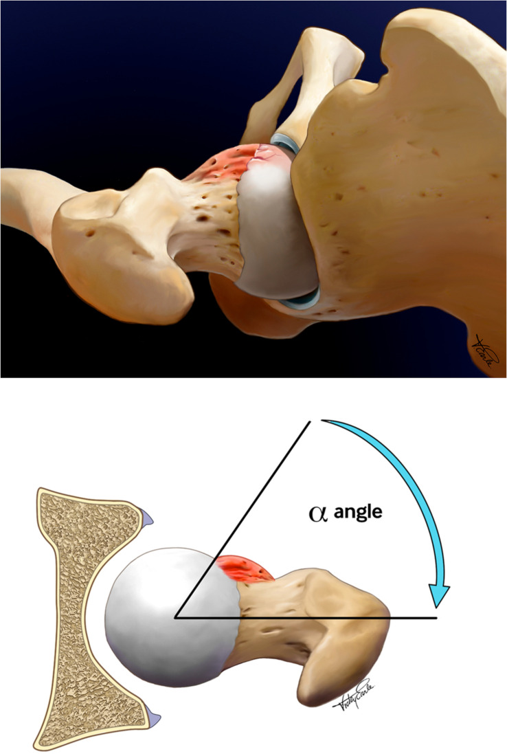
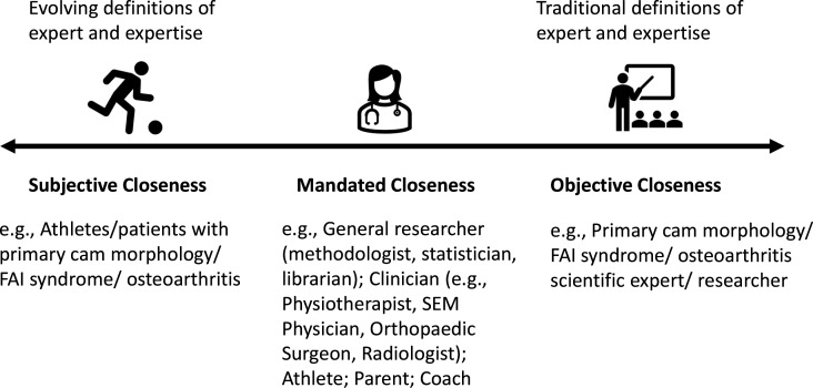
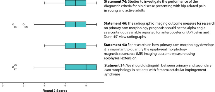
A diverse and inclusive Delphi panel (n=65 for rounds 1 and 2, representing 18 countries; 6 stakeholder groups; 40% women) agreed on 35 of 47 statements in 4 domains, while surfacing areas of tension and dissent. This Delphi panel agreed on four key issues essential to moving research and clinical care forward around primary cam morphology. They agreed on: (1) definition, confirming its conceptual attributes (tissue type, size, location, shape and ownership); (2) terminology-use 'morphology' and not terms with a negative connotation like 'lesion', 'abnormality' or 'deformity'; (3) taxonomy, distinguishing between primary and secondary cam morphology, and (4) imaging outcomes, a continuous bone/cartilage alpha angle on radial femoral head-neck MRI for primary cam morphology aetiology research.
This consensus provides athletes, patients, clinicians and researchers with a strong foundation to guide more precise communication, better clinical decision-making and higher value research about primary cam morphology and its natural history.
原发性凸轮形态是一种大多为良性的骨性隆起,发生于髋关节的股骨头 - 颈交界处,但在许多运动员群体中高度普遍。在一小部分并非良性的运动员中,由此导致的髋骨关节炎可能使人衰弱。临床医生、运动员、患者和研究人员在原发性凸轮形态的重要要素上尚未达成共识。我们旨在确定并提高在原发性凸轮形态定义、术语、分类法和影像学结果测量方面的共识水平。
为收集和汇总有见地的意见,一个专家小组——青年运动员髋关节研究协作组——通过在线德尔菲法对原发性凸轮形态定义、术语、分类法和影像学结果陈述进行评级,随后召开在线会议以探讨存在分歧和异议的领域。报告遵循《进行和报告德尔菲研究》。
一个多样化且具包容性的德尔菲小组(第1轮和第2轮有65人,代表18个国家;6个利益相关者群体;40%为女性)在4个领域的47项陈述中的35项上达成了一致,同时揭示了存在分歧和异议的领域。该德尔菲小组就推动围绕原发性凸轮形态的研究和临床护理向前发展至关重要的四个关键问题达成了一致。他们达成一致的是:(1)定义,确认其概念属性(组织类型、大小、位置、形状和所属);(2)术语——使用“形态”,而非带有负面含义的术语,如“病变”“异常”或“畸形”;(3)分类法,区分原发性和继发性凸轮形态;(4)影像学结果,用于原发性凸轮形态病因学研究的股骨头 - 颈桡侧MRI上连续的骨/软骨α角。
这一共识为运动员、患者、临床医生和研究人员提供了一个坚实的基础,以指导关于原发性凸轮形态及其自然史的更精确沟通、更好的临床决策和更具价值的研究。