Department of Bioengineering, Stanford University, Stanford, CA, USA.
Department of Chemistry and Chemical Biology, Harvard University, Cambridge, MA, USA.
Cell. 2023 Feb 2;186(3):543-559.e19. doi: 10.1016/j.cell.2022.12.035. Epub 2023 Jan 19.
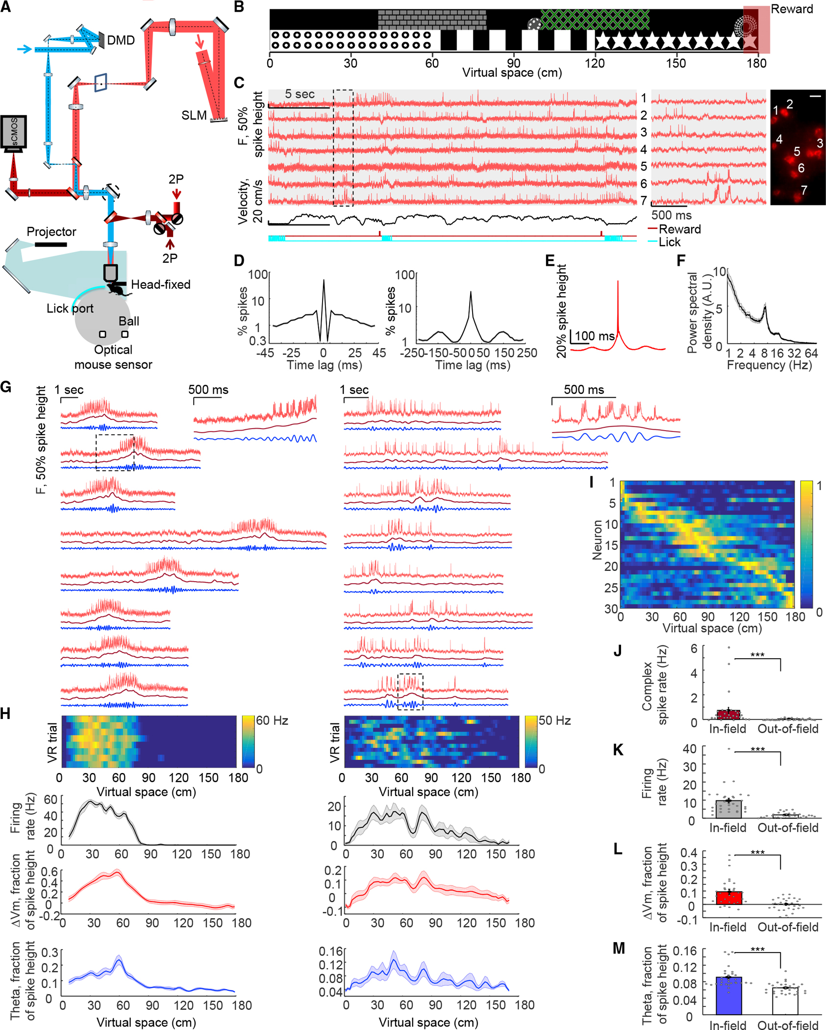
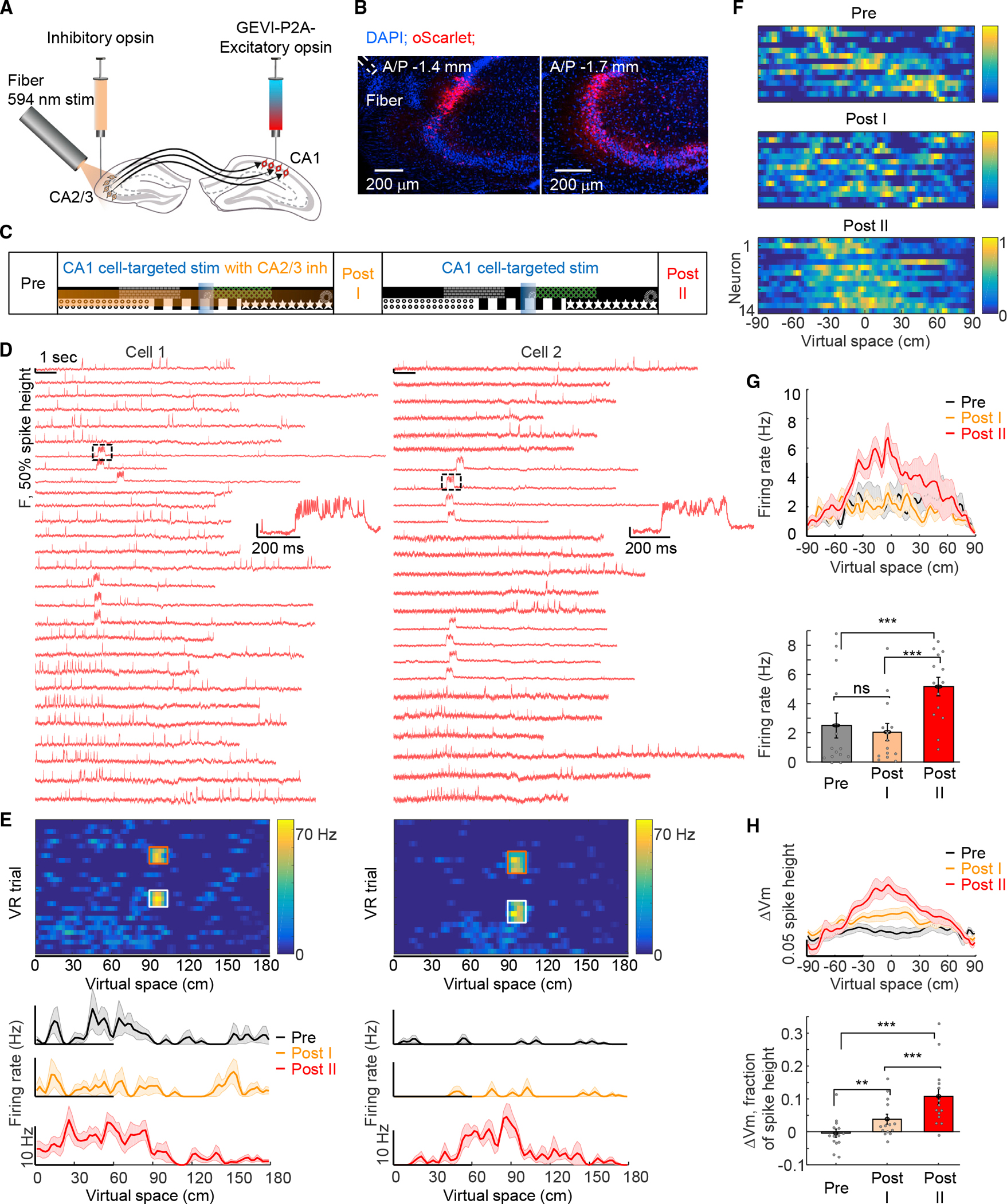
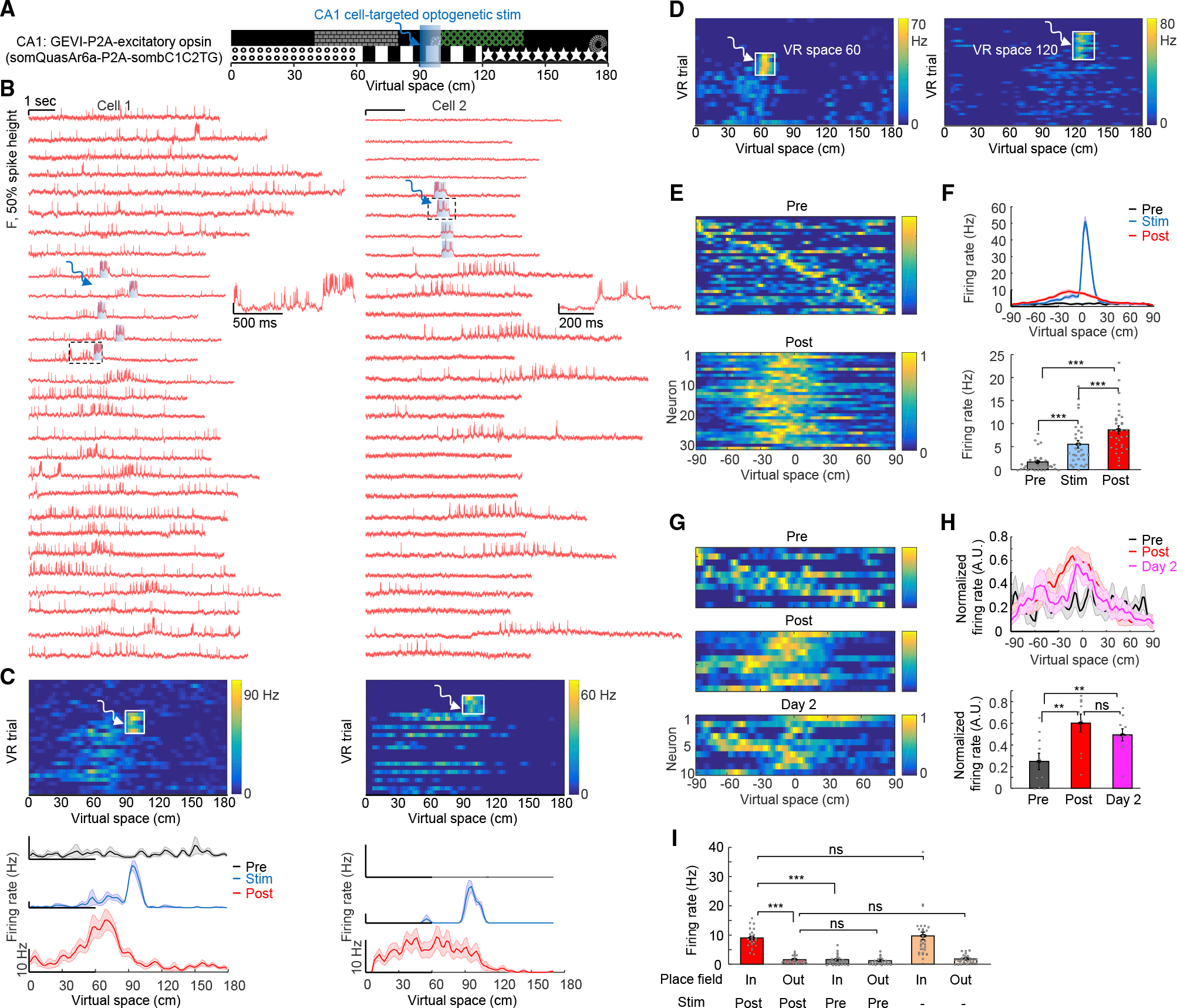
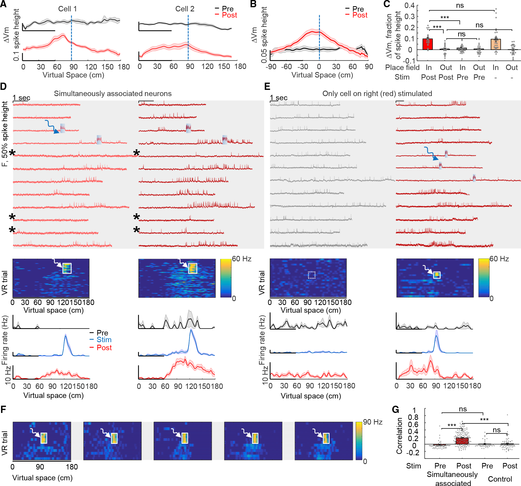
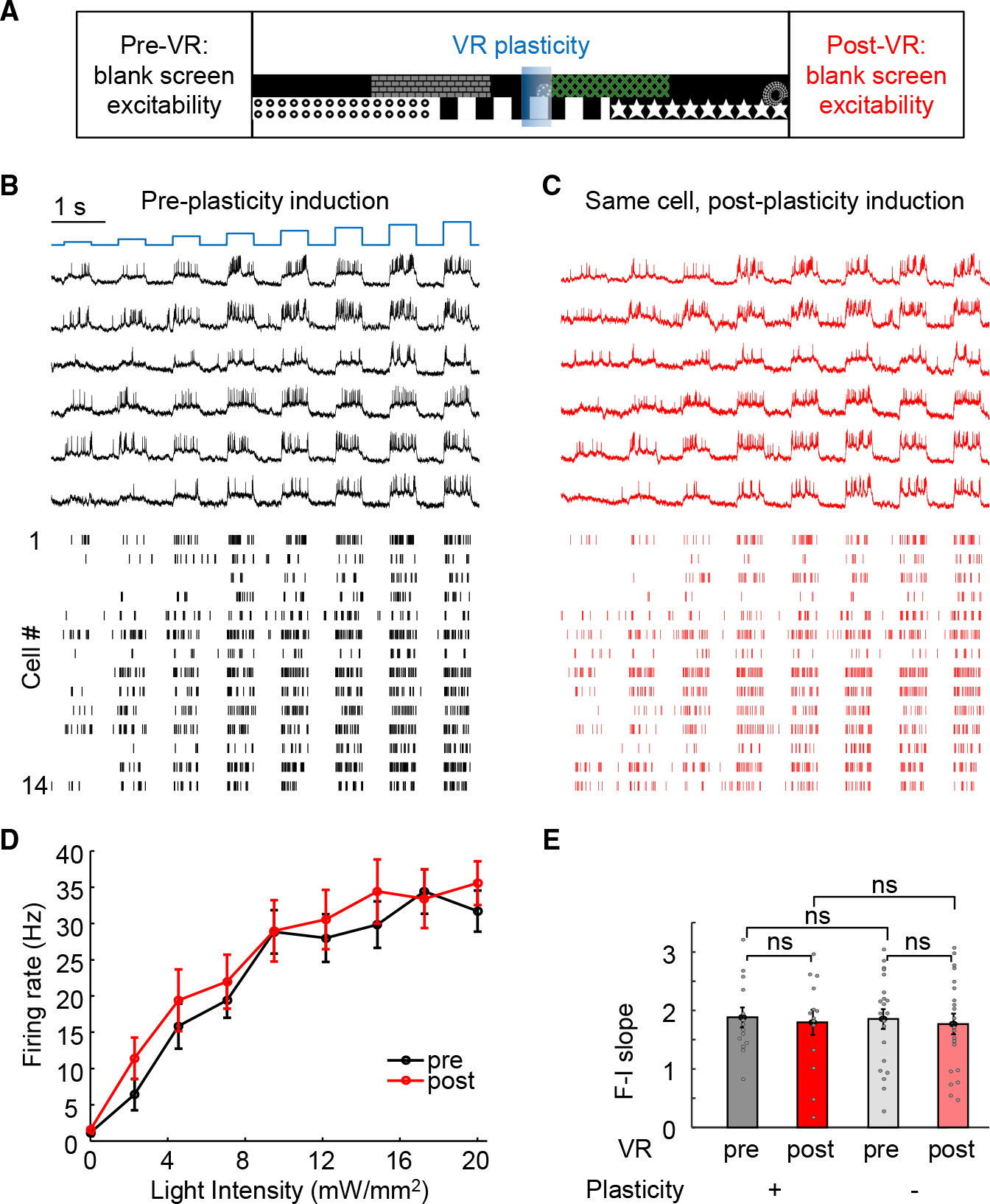
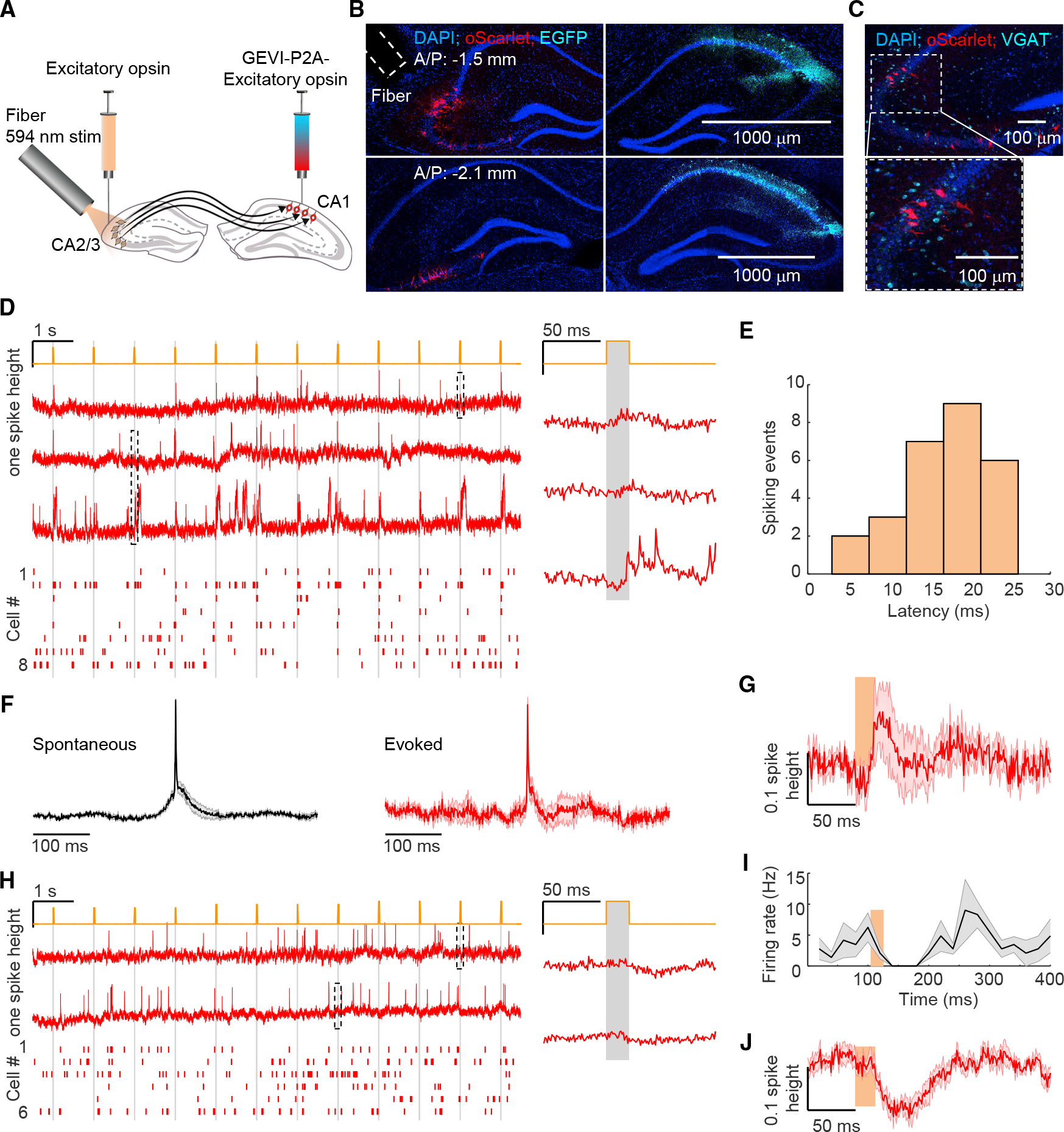
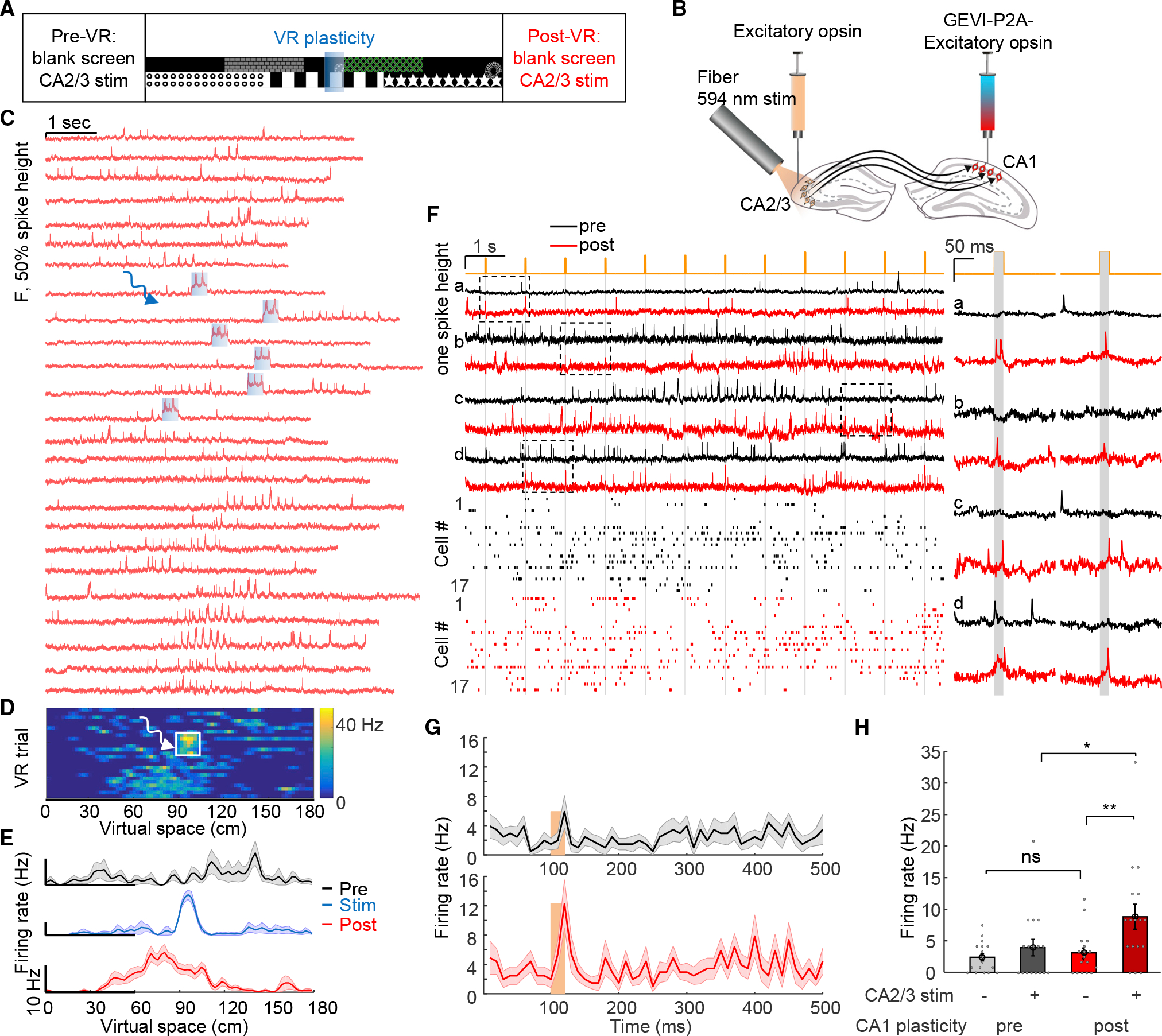
Learning has been associated with modifications of synaptic and circuit properties, but the precise changes storing information in mammals have remained largely unclear. We combined genetically targeted voltage imaging with targeted optogenetic activation and silencing of pre- and post-synaptic neurons to study the mechanisms underlying hippocampal behavioral timescale plasticity. In mice navigating a virtual-reality environment, targeted optogenetic activation of individual CA1 cells at specific places induced stable representations of these places in the targeted cells. Optical elicitation, recording, and modulation of synaptic transmission in behaving mice revealed that activity in presynaptic CA2/3 cells was required for the induction of plasticity in CA1 and, furthermore, that during induction of these place fields in single CA1 cells, synaptic input from CA2/3 onto these same cells was potentiated. These results reveal synaptic implementation of hippocampal behavioral timescale plasticity and define a methodology to resolve synaptic plasticity during learning and memory in behaving mammals.
学习与突触和电路性质的改变有关,但在哺乳动物中存储信息的确切变化在很大程度上仍不清楚。我们结合了基因靶向电压成像以及靶向光遗传学激活和抑制前突触和后突触神经元,以研究海马体行为时间尺度可塑性的机制。在导航虚拟现实环境的小鼠中,在特定位置靶向光遗传学激活单个 CA1 细胞,会在目标细胞中诱导这些位置的稳定表示。在行为小鼠中进行光学引发、记录和突触传递的调制,揭示了 CA2/3 细胞中的前突触活动对于 CA1 中的可塑性诱导是必需的,而且,在单个 CA1 细胞中诱导这些位置场期间,来自 CA2/3 的突触输入被增强到这些相同的细胞上。这些结果揭示了海马体行为时间尺度可塑性的突触实现,并定义了一种在行为哺乳动物中学习和记忆期间解析突触可塑性的方法。