Bhende Vishal V, Sharma Tanishq S, Sharma Ashwin S, Kumar Amit, Patel Nirja P, Majmudar Hardil P, Patel Mamta R, Patel Kruti A, Panesar Gurpreet, Soni Kunal, Dhami Kartik B, Pathan Sohilkhan R, Parmar Dushyant M, Nerurkar Paresh
Pediatric Cardiac Surgery, Bhanubhai and Madhuben Patel Cardiac Centre, Shree Krishna Hospital, Bhaikaka University, Karamsad, IND.
Pediatric Cardiac Surgery, Gujarat Cancer Society Medical College, Hospital and Research Centre, Ahmedabad, IND.
Cureus. 2023 Jan 18;15(1):e33942. doi: 10.7759/cureus.33942. eCollection 2023 Jan.
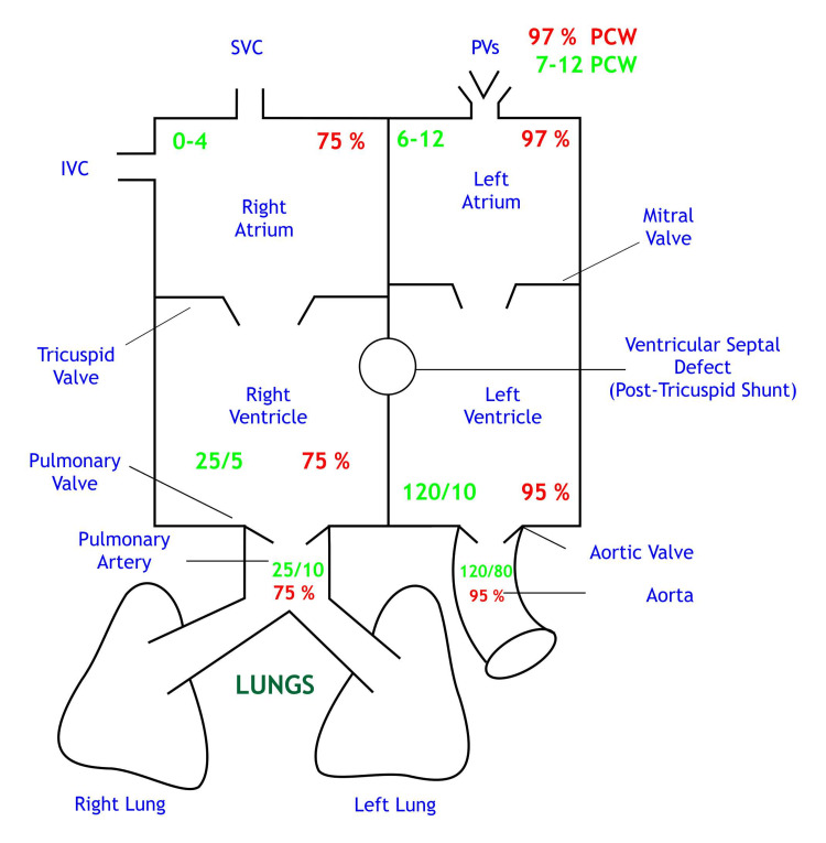
Background & aims Intracardiac shunts are abnormal channels of blood circulation within the heart that develop either as an additional blood flow pathway or as a replacement for the normal channels of blood circulation. They are the commonest types of congenital heart defects. Various methods are available in the present times to identify, localize or quantify left-to-right intracardiac shunts. Methods may vary in sensitivity, indicators, or types of equipment available. One such method used in almost all cardiac centers for a long time has been oximetry run to detect step-up differences in oxygen saturation values. In the oximetry run the main approach to detect and estimate the left-to-right (L-->R) shunts requires the oxygen concentration expressed as a proportion of saturation to be evaluated in blood samples which are obtained from the right atrium (RA) and pulmonary artery (PA), respectively. A left-to-right shunt can be considered if there is a significant increase (step-up) in blood saturation. A significant step-up is defined as a substantial rise in blood oxygen content or saturation that is higher than normal values. Methods Using a prospective observational design, this article investigates the application of the step-up method in detecting intracardiac shunts. The study was conducted between 2021 and 2022 on 35 pediatric cardiac patients (males/females, 24/11) diagnosed with post-tricuspid shunts. The pulmonary artery and right atrium were sampled before and after cardiopulmonary bypass surgery and analyzed using a blood gas test. As a result, nearly 91% of the patients had a saturation below 8%. However, the difference between PA oxygen saturation (SO & RASO before and after surgery was significant. As a result, the difference in O saturation helped detect the residual ventricular septal defect (VSD) after the surgery. Results There were no deaths or complications in this study. There were no re-interventions for post-tricuspid shunt surgery, though one patient had a step-up of >15% and residual VSD status was moderate to large on two-dimensional (2D) echocardiography. Conclusion A combination of physical findings, chest radiography, electrocardiogram (ECG), and echocardiography is routinely done for all these patients undergoing pediatric cardiac surgery. Echocardiography can detect the occurrence of shunt but does not calculate the shunt ratio. Transesophageal or epicardial echocardiography is the standard of care but has its limitations like perception difference between the operating surgeon and the person performing echocardiography. In this study, we have added an oximetry analysis of blood-gas samples before and after surgery and compared it to 2D echocardiography to test the validation of oximetry in isolation and comparison to 2D echocardiography.
背景与目的 心内分流是心脏内血液循环的异常通道,其形成方式要么是作为额外的血流途径,要么是作为正常血液循环通道的替代。它们是最常见的先天性心脏缺陷类型。目前有多种方法可用于识别、定位或量化左向右心内分流。这些方法在敏感性、指标或可用设备类型方面可能有所不同。长期以来,几乎所有心脏中心都使用的一种方法是进行血氧测定,以检测血氧饱和度值的升高差异。在血氧测定中,检测和估计左向右(L→R)分流的主要方法需要在分别从右心房(RA)和肺动脉(PA)采集的血样中评估以饱和度比例表示的氧浓度。如果血氧饱和度有显著升高(上升),则可认为存在左向右分流。显著上升定义为血氧含量或饱和度大幅升高且高于正常值。
方法 本文采用前瞻性观察设计,研究上升法在检测心内分流中的应用。该研究于2021年至2022年对35例诊断为三尖瓣后分流的儿科心脏病患者(男/女,24/11)进行。在体外循环手术前后对肺动脉和右心房进行采样,并使用血气测试进行分析。结果,近91%的患者饱和度低于8%。然而,手术前后肺动脉血氧饱和度(SO)与右心房血氧饱和度(RASO)之间的差异显著。因此,血氧饱和度的差异有助于检测手术后残留的室间隔缺损(VSD)。
结果 本研究中无死亡或并发症发生。三尖瓣后分流手术没有再次干预情况,尽管有1例患者上升超过15%,二维(2D)超声心动图显示残留VSD状态为中度至大量。
结论 对于所有接受儿科心脏手术的患者,通常会综合运用体格检查、胸部X线、心电图(ECG)和超声心动图。超声心动图可以检测分流的发生,但无法计算分流率。经食管或心外膜超声心动图是标准的检查方法,但存在局限性,如手术医生和进行超声心动图检查的人员之间的感知差异。在本研究中,我们增加了手术前后血气样本的血氧测定分析,并将其与二维超声心动图进行比较,以测试血氧测定单独使用时的有效性以及与二维超声心动图比较的结果。