Department of Brain Sciences, Imperial College London, London, UK.
UK Dementia Research Institute Centre for Care Research and Technology at Imperial College London, London, UK.
Alzheimers Dement. 2023 Jul;19(7):3065-3077. doi: 10.1002/alz.12934. Epub 2023 Jan 25.
Traumatic brain injury (TBI) is a dementia risk factor, with Alzheimer's disease (AD) more common following injury. Patterns of neurodegeneration produced by TBI can be compared to AD and aging using volumetric MRI.
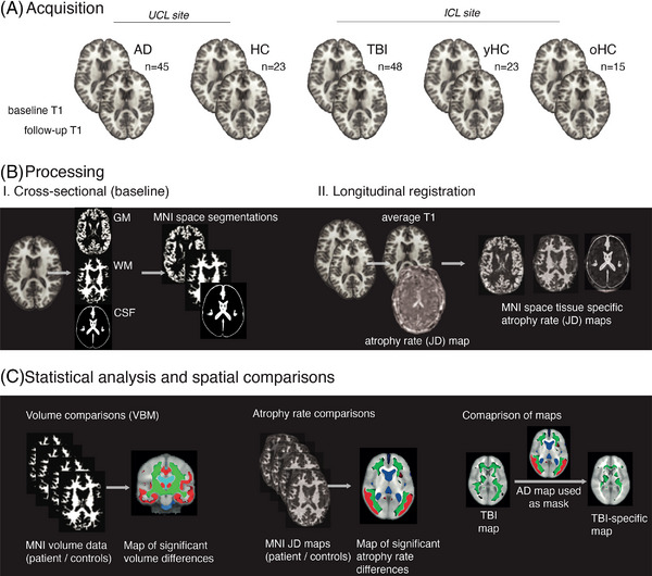
A total of 55 patients after moderate to severe TBI (median age 40), 45 with AD (median age 69), and 61 healthy volunteers underwent magnetic resonance imaging over 2 years. Atrophy patterns were compared.
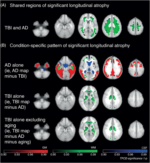
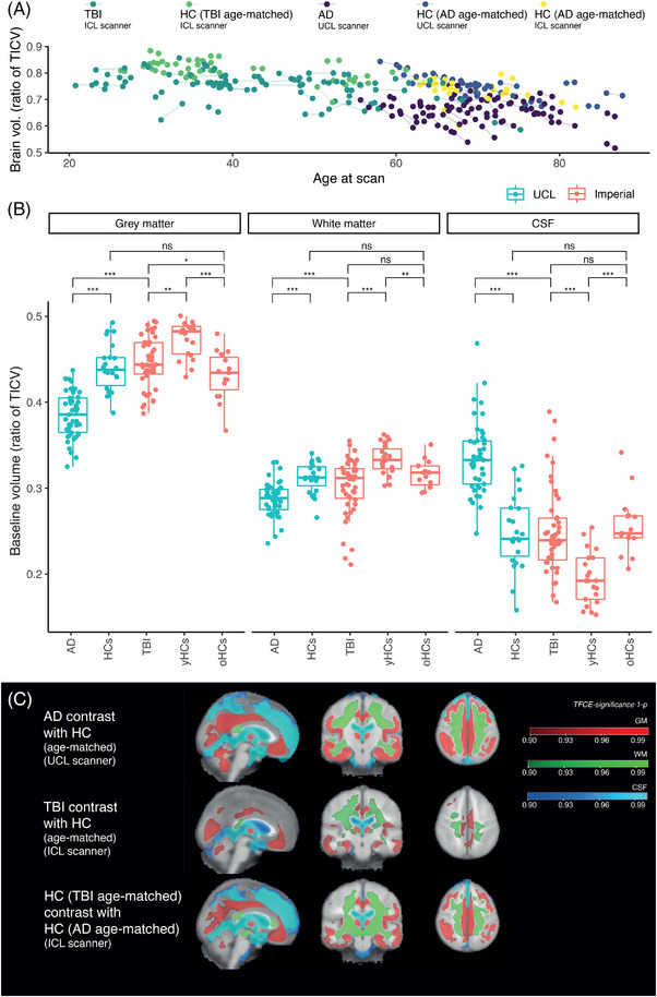
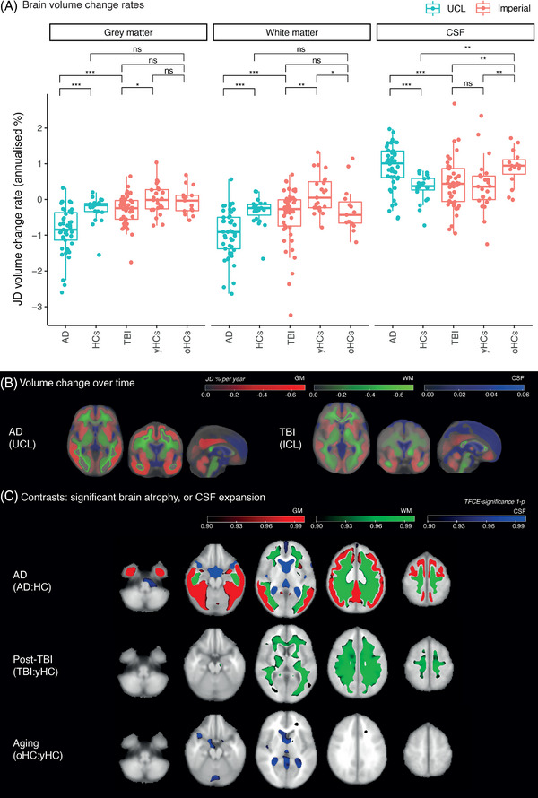
AD patients had markedly lower baseline volumes. TBI was associated with increased white matter (WM) atrophy, particularly involving corticospinal tracts and callosum, whereas AD rates were increased across white and gray matter (GM). Subcortical WM loss was shared in AD/TBI, but deep WM atrophy was TBI-specific and cortical atrophy AD-specific. Post-TBI atrophy patterns were distinct from aging, which resembled AD.
Post-traumatic neurodegeneration 1.9-4.0 years (median) following moderate-severe TBI is distinct from aging/AD, predominantly involving central WM. This likely reflects distributions of axonal injury, a neurodegeneration trigger.
We compared patterns of brain atrophy longitudinally after moderate to severe TBI in late-onset AD and healthy aging. Patients after TBI had abnormal brain atrophy involving the corpus callosum and other WM tracts, including corticospinal tracts, in a pattern that was specific and distinct from AD and aging. This pattern is reminiscent of axonal injury following TBI, and atrophy rates were predicted by the extent of axonal injury on diffusion tensor imaging, supporting a relationship between early axonal damage and chronic neurodegeneration.
创伤性脑损伤(TBI)是痴呆的一个风险因素,受伤后更常发生阿尔茨海默病(AD)。使用容积 MRI 可以将 TBI 引起的神经退行性变模式与 AD 和衰老进行比较。
共有 55 名中重度 TBI 后患者(中位数年龄 40 岁)、45 名 AD 患者(中位数年龄 69 岁)和 61 名健康志愿者在 2 年内接受了磁共振成像检查。比较了萎缩模式。
AD 患者的基线体积明显较低。TBI 与白质(WM)萎缩增加有关,特别是涉及皮质脊髓束和胼胝体,而 AD 率则增加了白质和灰质(GM)。AD/TBI 存在皮质下 WM 丢失,但深部 WM 萎缩是 TBI 特异性的,皮质萎缩是 AD 特异性的。TBI 后的萎缩模式与衰老不同,与 AD 相似。
中重度 TBI 后 1.9-4.0 年(中位数)的创伤后神经退行性变与衰老/AD 不同,主要涉及中央 WM。这可能反映了轴突损伤的分布,这是一种神经退行性变的触发因素。
我们比较了中重度 TBI 后迟发性 AD 和健康衰老患者的大脑萎缩模式。TBI 后患者的大脑萎缩模式异常,涉及胼胝体和其他 WM 束,包括皮质脊髓束,这种模式与 AD 和衰老不同且具有特异性。这种模式让人联想到 TBI 后的轴突损伤,萎缩率可通过弥散张量成像上的轴突损伤程度预测,支持早期轴突损伤与慢性神经退行性变之间的关系。