Liu Kunxiang, Liu Bo, Zhang Yuhong, Wu Qinian, Zhong Ming, Shang Lindong, Wang Yu, Liang Peng, Wang Weiguo, Zhao Qi, Li Bei
State Key Laboratory of Applied Optics, Changchun Institute of Optics, Fine Mechanics and Physics, Chinese Academy of Sciences, Changchun 130033, PR China.
University of Chinese Academy of Sciences, Beijing 100049, PR China.
Comput Struct Biotechnol J. 2022 Dec 30;21:802-811. doi: 10.1016/j.csbj.2022.12.050. eCollection 2023.
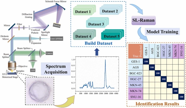
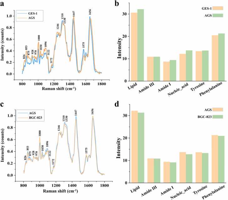
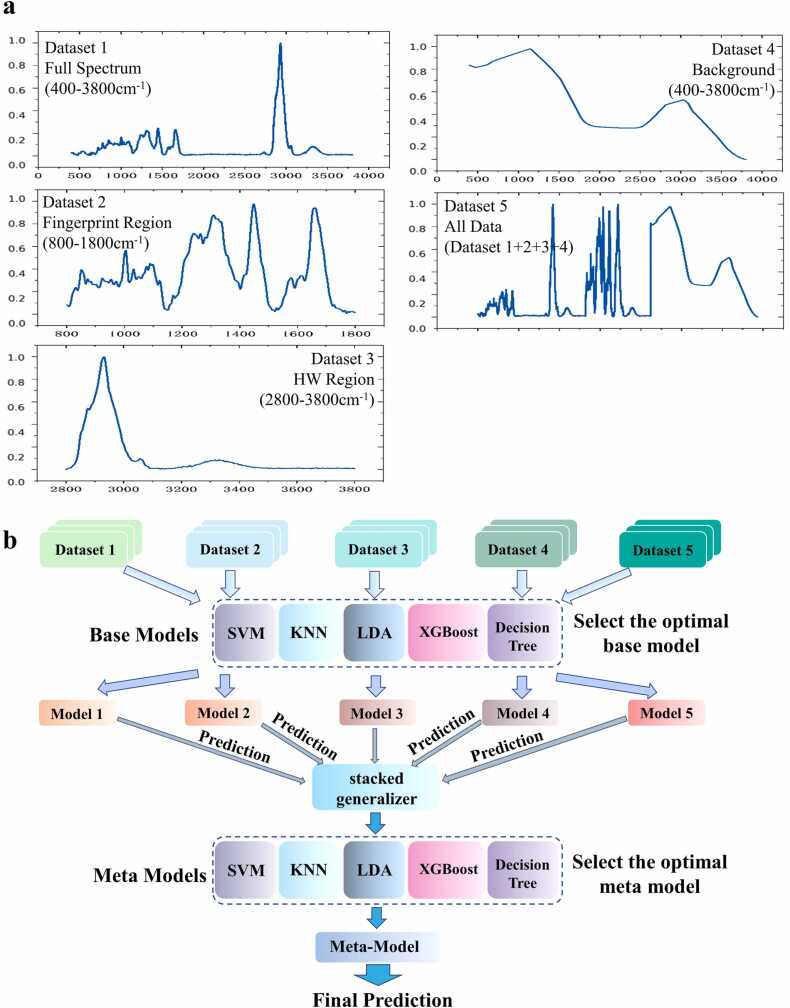
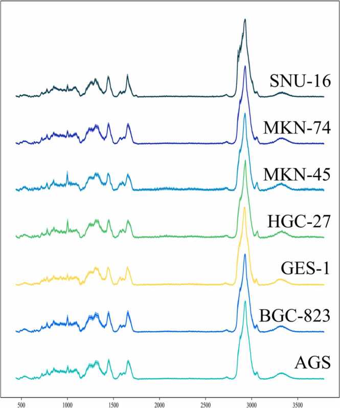
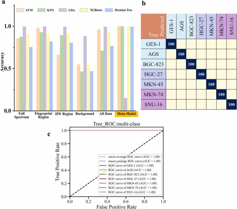
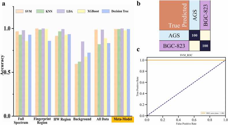
Cell misuse and cross-contamination can affect the accuracy of cell research results and result in wasted time, manpower and material resources. Thus, cell line identification is important and necessary. At present, the commonly used cell line identification methods need cell staining and culturing. There is therefore a need to develop a new method for the rapid and automated identification of cell lines. Raman spectroscopy has become one of the emerging techniques in the field of microbial identification, with the advantages of being rapid and noninvasive and providing molecular information for biological samples, which is beneficial in the identification of cell lines. In this study, we built a library of Raman spectra for gastric mucosal epithelial cell lines GES-1 and gastric cancer cell lines, such as AGS, BGC-823, HGC-27, MKN-45, MKN-74 and SNU-16. Five spectral datasets were constructed using spectral data and included the full spectrum, fingerprint region, high-wavelength number region and Raman background of Raman spectra. A stacking ensemble learning model, SL-Raman, was built for different datasets, and gastric cancer cell identification was achieved. For the gastric cancer cells we studied, the differentiation accuracy of SL-Raman was 100% for one of the gastric cancer cells and 100% for six of the gastric cancer cells. Additionally, the separation accuracy for two gastric cancer cells with different degrees of differentiation was 100%. These results demonstrate that Raman spectroscopy combined with SL-Raman may be a new method for the rapid and accurate identification of gastric cancer. In addition, the accuracy of 94.38% for classifying Raman spectral background data using machine learning demonstrates that the Raman spectral background contains some useful spectral features. These data have been overlooked in previous studies.
细胞的误用和交叉污染会影响细胞研究结果的准确性,导致时间、人力和物力的浪费。因此,细胞系鉴定非常重要且必要。目前,常用的细胞系鉴定方法需要细胞染色和培养。因此,需要开发一种新的细胞系快速自动鉴定方法。拉曼光谱已成为微生物鉴定领域新兴的技术之一,具有快速、无创的优点,并能为生物样品提供分子信息,这有利于细胞系的鉴定。在本研究中,我们构建了胃黏膜上皮细胞系GES-1和胃癌细胞系(如AGS、BGC-823、HGC-27、MKN-45、MKN-74和SNU-16)的拉曼光谱库。利用光谱数据构建了五个光谱数据集,包括拉曼光谱的全谱、指纹区、高波数区和拉曼背景。针对不同数据集构建了堆叠集成学习模型SL-Raman,并实现了胃癌细胞的鉴定。对于我们研究的胃癌细胞,SL-Raman对其中一种胃癌细胞的鉴别准确率为100%,对六种胃癌细胞的鉴别准确率为100%。此外,对两种不同分化程度的胃癌细胞的分离准确率为100%。这些结果表明,拉曼光谱结合SL-Raman可能是一种快速准确鉴定胃癌的新方法。此外,利用机器学习对拉曼光谱背景数据进行分类的准确率为94.38%,表明拉曼光谱背景包含一些有用的光谱特征。这些数据在以往的研究中被忽视了。