• 文献检索
  • 文档翻译
  • 深度研究
  • 学术资讯
  • Suppr Zotero 插件Zotero 插件
  • 邀请有礼
  • 套餐&价格
  • 历史记录
应用&插件
Suppr Zotero 插件Zotero 插件浏览器插件Mac 客户端Windows 客户端微信小程序
定价
高级版会员购买积分包购买API积分包
服务
文献检索文档翻译深度研究API 文档MCP 服务
关于我们
关于 Suppr公司介绍联系我们用户协议隐私条款
关注我们

Suppr 超能文献

核心技术专利:CN118964589B侵权必究
粤ICP备2023148730 号-1Suppr @ 2026

文献检索

告别复杂PubMed语法,用中文像聊天一样搜索,搜遍4000万医学文献。AI智能推荐,让科研检索更轻松。

立即免费搜索

文件翻译

保留排版,准确专业,支持PDF/Word/PPT等文件格式,支持 12+语言互译。

免费翻译文档

深度研究

AI帮你快速写综述,25分钟生成高质量综述,智能提取关键信息,辅助科研写作。

立即免费体验

蛛网膜下腔出血后与脑缺血相关的脑血管痉挛的早期诊断:计算机断层扫描灌注和经颅多普勒作为准确方法的评估

Early diagnosis of cerebral vasospasm associated with cerebral ischemia following subarachnoid hemorrhage: Evaluation of computed tomography perfusion and transcranial doppler as accurate methods.

作者信息

Tsolaki Vasiliki, Aravantinou-Fatorou Aikaterini, Georgakopoulou Vasiliki Epameinondas, Spandidos Demetrios A, Papalexis Petros, Mathioudakis Nikolaos, Tarantinos Kyriakos, Trakas Nikolaos, Sklapani Pagona, Fotakopoulos George

机构信息

Department of Pulmonary and Critical Care Medicine, General University Hospital of Larisa, 41221 Larisa, Greece.

First Department of Internal Medicine, Laiko General Hospital, Medical School, National and Kapodistrian University of Athens, 11527 Athens, Greece.

出版信息

Med Int (Lond). 2022 Nov 2;2(6):34. doi: 10.3892/mi.2022.59. eCollection 2022 Nov-Dec.

DOI:10.3892/mi.2022.59
PMID:36699155
原文链接:https://pmc.ncbi.nlm.nih.gov/articles/PMC9829237/
Abstract

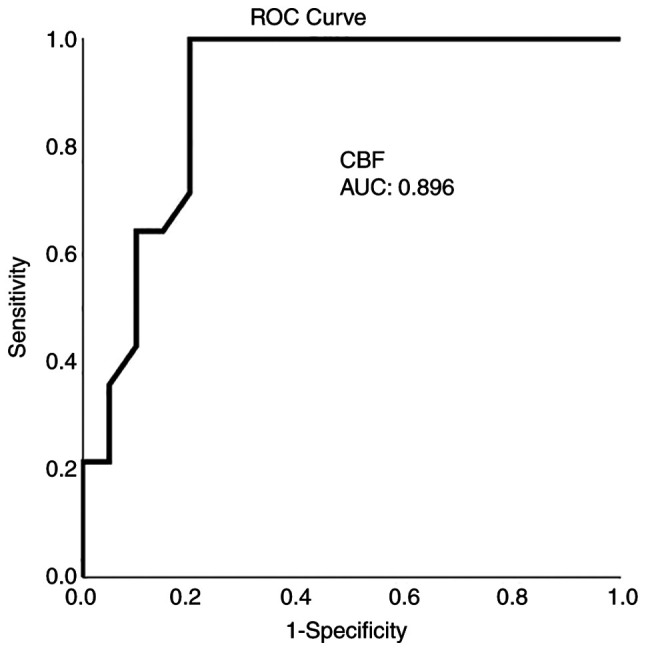
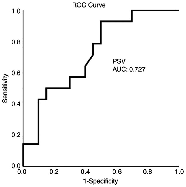
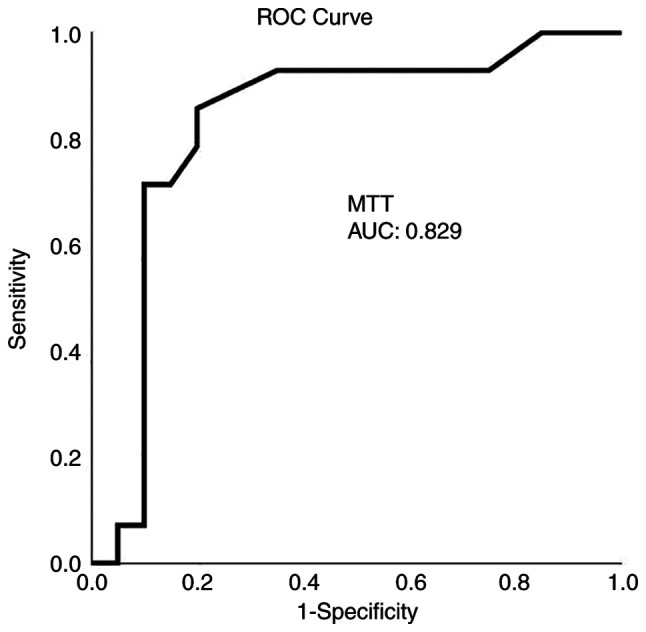
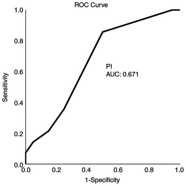
Cerebral vasospasm (CV) constitutes a major post-operative complication and source of morbidity in cases of subarachnoid hemorrhage (SAH). The early detection of CV in SAH may be difficult both clinically and radiographically. The present pilot study thus aimed to evaluate the practicability of the technique in a tertiary healthcare setting and to assess the diagnostic effectiveness of various diagnostic computed tomography (CT) perfusion (CTP) aspects in predicting the clinical outcome of patients with SAH (traumatic and aneurysmal). A retrospective study including 34 patients in a tertiary care hospital was thus conducted. The results revealed that of the 34 patients, 18 (52.9%) were males, and the mean age was 54.4±18.5 years (16-85 years old; range, 69 years). In total, 15 (44.1%) patients had traumatic SAH following traumatic brain injury (TBI), 11 (33.3%) had aneurysmal SAH, and 8 patients (23.6%) presented with TBI without SAH as controls. CTP was performed on the third to the sixth day, and 15-20 min prior to CPT, a transcranial Doppler ultrasound was performed. Clinical outcomes were documented at 30 days using a CT scan and a complete neurological evaluation, including Glasgow Coma Scale assessment. The results of a multivariate analysis revealed that cerebral blood flow (CBF) was an independent factor for detecting an ischemic event in 1 month (P=0.003). On the whole, the present study demonstrates that CTP, and consequently CBF, is a considerable index that may identify the onset of cerebral ischemia in patients with SAH.

摘要

脑血管痉挛(CV)是蛛网膜下腔出血(SAH)病例中的一种主要术后并发症和发病源。SAH中CV的早期检测在临床和影像学上都可能很困难。因此,本初步研究旨在评估该技术在三级医疗环境中的实用性,并评估各种诊断性计算机断层扫描(CT)灌注(CTP)方面在预测SAH(创伤性和动脉瘤性)患者临床结局方面的诊断有效性。因此,在一家三级护理医院进行了一项包括34例患者的回顾性研究。结果显示,在这34例患者中,18例(52.9%)为男性,平均年龄为54.4±18.5岁(16 - 85岁;范围为69岁)。总共有15例(44.1%)患者在创伤性脑损伤(TBI)后发生创伤性SAH,11例(33.3%)有动脉瘤性SAH,8例患者(23.6%)表现为TBI但无SAH作为对照。在第3至6天进行CTP检查,在CTP检查前15 - 20分钟进行经颅多普勒超声检查。使用CT扫描和包括格拉斯哥昏迷量表评估在内的完整神经学评估在30天时记录临床结局。多变量分析结果显示,脑血流量(CBF)是检测1个月内缺血事件的独立因素(P = 0.003)。总体而言,本研究表明CTP,进而CBF,是一个重要指标,可能识别SAH患者脑缺血的发作。

https://cdn.ncbi.nlm.nih.gov/pmc/blobs/3552/9829237/566115d50523/mi-02-06-00059-g03.jpg
https://cdn.ncbi.nlm.nih.gov/pmc/blobs/3552/9829237/e6fc9de5a96a/mi-02-06-00059-g00.jpg
https://cdn.ncbi.nlm.nih.gov/pmc/blobs/3552/9829237/7e8090b59243/mi-02-06-00059-g01.jpg
https://cdn.ncbi.nlm.nih.gov/pmc/blobs/3552/9829237/faa8e90c4c49/mi-02-06-00059-g02.jpg
https://cdn.ncbi.nlm.nih.gov/pmc/blobs/3552/9829237/566115d50523/mi-02-06-00059-g03.jpg

相似文献

1
Early diagnosis of cerebral vasospasm associated with cerebral ischemia following subarachnoid hemorrhage: Evaluation of computed tomography perfusion and transcranial doppler as accurate methods.蛛网膜下腔出血后与脑缺血相关的脑血管痉挛的早期诊断:计算机断层扫描灌注和经颅多普勒作为准确方法的评估
Med Int (Lond). 2022 Nov 2;2(6):34. doi: 10.3892/mi.2022.59. eCollection 2022 Nov-Dec.
2
Cost-effectiveness of CT angiography and perfusion imaging for delayed cerebral ischemia and vasospasm in aneurysmal subarachnoid hemorrhage.CT血管造影和灌注成像对动脉瘤性蛛网膜下腔出血后迟发性脑缺血和血管痉挛的成本效益分析
AJNR Am J Neuroradiol. 2014 Sep;35(9):1714-20. doi: 10.3174/ajnr.A3947. Epub 2014 May 8.
3
Traumatic brain injury and intracranial hemorrhage-induced cerebral vasospasm: a systematic review.创伤性脑损伤和颅内出血引起的脑血管痉挛:系统评价。
Neurosurg Focus. 2017 Nov;43(5):E14. doi: 10.3171/2017.8.FOCUS17431.
4
Preventive effect of continuous cisternal irrigation with magnesium sulfate solution on angiographic cerebral vasospasms associated with aneurysmal subarachnoid hemorrhages: a randomized controlled trial.硫酸镁溶液持续脑池灌洗对动脉瘤性蛛网膜下腔出血相关脑血管造影血管痉挛的预防作用:一项随机对照试验。
J Neurosurg. 2016 Jan;124(1):18-26. doi: 10.3171/2015.1.JNS142757. Epub 2015 Jul 31.
5
Early whole-brain CT perfusion for detection of patients at risk for delayed cerebral ischemia after subarachnoid hemorrhage.早期全脑 CT 灌注检测蛛网膜下腔出血后迟发性脑缺血高危患者。
J Neurosurg. 2016 Jul;125(1):128-36. doi: 10.3171/2015.6.JNS15720. Epub 2015 Dec 18.
6
Molsidomine for the prevention of vasospasm-related delayed ischemic neurological deficits and delayed brain infarction and the improvement of clinical outcome after subarachnoid hemorrhage: a single-center clinical observational study.莫西赛利预防蛛网膜下腔出血后血管痉挛相关的迟发性缺血性神经功能缺损和迟发性脑梗死及改善临床结局:一项单中心临床观察性研究
J Neurosurg. 2016 Jan;124(1):51-8. doi: 10.3171/2014.12.JNS13846. Epub 2015 Jul 10.
7
The value of computed tomography perfusion & transcranial Doppler in early diagnosis of cerebral vasospasm in aneurysmal & traumatic subarachnoid hemorrhage.计算机断层扫描灌注成像及经颅多普勒在动脉瘤性和创伤性蛛网膜下腔出血后脑血管痉挛早期诊断中的价值
Future Sci OA. 2018 Jun 26;4(6):FSO313. doi: 10.4155/fsoa-2018-0015. eCollection 2018 Jul.
8
Whole brain CT perfusion combined with CT angiography in patients with subarachnoid hemorrhage and cerebral vasospasm.全脑CT灌注联合CT血管造影在蛛网膜下腔出血和脑血管痉挛患者中的应用
Clin Neurol Neurosurg. 2013 Dec;115(12):2496-501. doi: 10.1016/j.clineuro.2013.10.004. Epub 2013 Oct 16.
9
Value of sildenafil treatment for the prevention of vasospasm‑related delayed ischemic neurological deficits and delayed brain infarction following aneurysmal subarachnoid hemorrhage.西地那非治疗对预防动脉瘤性蛛网膜下腔出血后血管痉挛相关的迟发性缺血性神经功能缺损和迟发性脑梗死的价值。
Med Int (Lond). 2023 Mar 20;3(2):19. doi: 10.3892/mi.2023.79. eCollection 2023 Mar-Apr.
10
Posttraumatic cerebral arterial spasm: transcranial Doppler ultrasound, cerebral blood flow, and angiographic findings.创伤后脑动脉痉挛:经颅多普勒超声、脑血流量及血管造影结果
J Neurosurg. 1992 Oct;77(4):575-83. doi: 10.3171/jns.1992.77.4.0575.

引用本文的文献

1
Long-term outcomes following the clipping of ruptured intracranial aneurysms of the anterior circulation: A retrospective institutional study.前循环破裂颅内动脉瘤夹闭术后的长期预后:一项机构回顾性研究。
Med Int (Lond). 2025 Jul 31;5(5):58. doi: 10.3892/mi.2025.257. eCollection 2025 Sep-Oct.
2
Treatment Outcomes of Clazosentan Use During the Perioperative Period for Subarachnoid Hemorrhage.蛛网膜下腔出血围手术期使用氯沙坦的治疗结果
Cureus. 2025 Feb 23;17(2):e79497. doi: 10.7759/cureus.79497. eCollection 2025 Feb.
3
Microsurgery Treatment as an Optimal Management of Posterior Cerebral Artery Aneurysms: A Systematic Review and Meta-Analysis.

本文引用的文献

1
The value of computed tomography perfusion & transcranial Doppler in early diagnosis of cerebral vasospasm in aneurysmal & traumatic subarachnoid hemorrhage.计算机断层扫描灌注成像及经颅多普勒在动脉瘤性和创伤性蛛网膜下腔出血后脑血管痉挛早期诊断中的价值
Future Sci OA. 2018 Jun 26;4(6):FSO313. doi: 10.4155/fsoa-2018-0015. eCollection 2018 Jul.
2
Management of delayed cerebral ischemia after subarachnoid hemorrhage.蛛网膜下腔出血后迟发性脑缺血的管理
Crit Care. 2016 Oct 14;20(1):277. doi: 10.1186/s13054-016-1447-6.
3
CT perfusion and delayed cerebral ischemia in aneurysmal subarachnoid hemorrhage: a systematic review and meta-analysis.
显微外科手术治疗作为大脑后动脉动脉瘤的最佳治疗方法:一项系统评价和荟萃分析
Cureus. 2025 Jan 22;17(1):e77856. doi: 10.7759/cureus.77856. eCollection 2025 Jan.
4
The Outcome after Surgical Management of Ruptured Versus Unruptured Anterior Circulation Aneurysms.破裂与未破裂的前循环动脉瘤手术治疗后的结果
Maedica (Bucur). 2024 Jun;19(2):221-228. doi: 10.26574/maedica.2024.19.2.221.
5
Comparison of efficacy between clazosentan and fasudil hydrochloride-based management of vasospasm after subarachnoid hemorrhage focusing on older and WFNS grade V patients: a single-center experience in Japan.比较氯苯唑酸和盐酸法舒地尔在蛛网膜下腔出血后血管痉挛治疗中的疗效:日本单中心经验,重点关注老年和 WFNS 分级 V 患者。
Neurosurg Rev. 2024 Mar 13;47(1):113. doi: 10.1007/s10143-024-02345-9.
6
Effect of circulating miR‑126 levels on intracranial aneurysms and their predictive value for the rupture of aneurysms: A systematic review and meta‑analysis.循环miR-126水平对颅内动脉瘤的影响及其对动脉瘤破裂的预测价值:一项系统评价和荟萃分析
Exp Ther Med. 2023 Jul 10;26(2):411. doi: 10.3892/etm.2023.12110. eCollection 2023 Aug.
CT 灌注与动脉瘤性蛛网膜下腔出血后迟发性脑缺血:系统评价和荟萃分析。
J Cereb Blood Flow Metab. 2014 Feb;34(2):200-7. doi: 10.1038/jcbfm.2013.208. Epub 2013 Nov 27.
4
Prediction of poststroke hemorrhagic transformation using computed tomography perfusion.基于 CT 灌注成像预测卒中后出血转化。
Stroke. 2013 Nov;44(11):3039-43. doi: 10.1161/STROKEAHA.113.002396. Epub 2013 Sep 3.
5
Value of transcranial Doppler, perfusion-CT and neurological evaluation to forecast secondary ischemia after aneurysmal SAH.经颅多普勒、灌注CT及神经学评估对预测动脉瘤性蛛网膜下腔出血后继发性缺血的价值。
Neurocrit Care. 2014 Jun;20(3):406-12. doi: 10.1007/s12028-013-9896-0.
6
Acute stroke imaging research roadmap II and international survey of acute stroke imaging capabilities: we need your help!急性卒中成像研究路线图II及急性卒中成像能力国际调查:我们需要您的帮助!
AJNR Am J Neuroradiol. 2013 Sep;34(9):1671. doi: 10.3174/ajnr.A3698. Epub 2013 Jul 18.
7
Value of Perfusion CT, Transcranial Doppler Sonography, and Neurological Examination to Detect Delayed Vasospasm after Aneurysmal Subarachnoid Hemorrhage.灌注CT、经颅多普勒超声及神经学检查在检测动脉瘤性蛛网膜下腔出血后迟发性血管痉挛中的价值
Radiol Res Pract. 2012;2012:231206. doi: 10.1155/2012/231206. Epub 2012 Sep 24.
8
Using quantitative CT perfusion for evaluation of delayed cerebral ischemia following aneurysmal subarachnoid hemorrhage.采用定量 CT 灌注评估颅内动脉瘤性蛛网膜下腔出血后迟发性脑缺血。
AJNR Am J Neuroradiol. 2011 Dec;32(11):2047-53. doi: 10.3174/ajnr.A2693. Epub 2011 Sep 29.
9
Detection and monitoring of vasospasm and delayed cerebral ischemia: a review and assessment of the literature.血管痉挛和迟发性脑缺血的检测和监测:文献回顾与评估。
Neurocrit Care. 2011 Sep;15(2):312-7. doi: 10.1007/s12028-011-9594-8.
10
Vasospasm after subarachnoid hemorrhage: a 3D rotational angiography study.蛛网膜下腔出血后的血管痉挛:一项三维旋转血管造影研究。
Acta Neurochir Suppl. 2011;110(Pt 2):221-5. doi: 10.1007/978-3-7091-0356-2_40.