• 文献检索
  • 文档翻译
  • 深度研究
  • 学术资讯
  • Suppr Zotero 插件Zotero 插件
  • 邀请有礼
  • 套餐&价格
  • 历史记录
应用&插件
Suppr Zotero 插件Zotero 插件浏览器插件Mac 客户端Windows 客户端微信小程序
定价
高级版会员购买积分包购买API积分包
服务
文献检索文档翻译深度研究API 文档MCP 服务
关于我们
关于 Suppr公司介绍联系我们用户协议隐私条款
关注我们

Suppr 超能文献

核心技术专利:CN118964589B侵权必究
粤ICP备2023148730 号-1Suppr @ 2026

文献检索

告别复杂PubMed语法,用中文像聊天一样搜索,搜遍4000万医学文献。AI智能推荐,让科研检索更轻松。

立即免费搜索

文件翻译

保留排版,准确专业,支持PDF/Word/PPT等文件格式,支持 12+语言互译。

免费翻译文档

深度研究

AI帮你快速写综述,25分钟生成高质量综述,智能提取关键信息,辅助科研写作。

立即免费体验

使用自监督等变注意力机制对胸部X光片上的肺部异常进行定位

Localization of lung abnormalities on chest X-rays using self-supervised equivariant attention.

作者信息

D'Souza Gavin, Reddy N V Subba, Manjunath K N

机构信息

Department of Instrumentation and Control Engineering, Manipal Institute of Technology, Manipal Academy of Higher Education, Manipal, Karnataka 576104 India.

Department of Information Technology, Manipal Institute of Technology Bengaluru, Manipal Academy of Higher Education, Manipal, Karnataka 560064 India.

出版信息

Biomed Eng Lett. 2022 Nov 3;13(1):21-30. doi: 10.1007/s13534-022-00249-5. eCollection 2023 Feb.

DOI:10.1007/s13534-022-00249-5
PMID:36711159
原文链接:https://pmc.ncbi.nlm.nih.gov/articles/PMC9873849/
Abstract

UNLABELLED

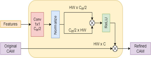
Chest X-Ray (CXR) images provide most anatomical details and the abnormalities on a 2D plane. Therefore, a 2D view of the 3D anatomy is sometimes sufficient for the initial diagnosis. However, close to fourteen commonly occurring diseases are sometimes difficult to identify by visually inspecting the images. Therefore, there is a drift toward developing computer-aided assistive systems to help radiologists. This paper proposes a deep learning model for the classification and localization of chest diseases by using image-level annotations. The model consists of a modified Resnet50 backbone for extracting feature corpus from the images, a classifier, and a pixel correlation module (PCM). During PCM training, the network is a weight-shared siamese architecture where the first branch applies the affine transform to the image before feeding to the network, while the second applies the same transform to the network output. The method was evaluated on CXR from the clinical center in the ratio of 70:20 for training and testing. The model was developed and tested using the cloud computing platform Google Colaboratory (NVidia Tesla P100 GPU, 16 GB of RAM). A radiologist subjectively validated the results. Our model trained with the configurations mentioned in this paper outperformed benchmark results.

SUPPLEMENTARY INFORMATION

The online version contains supplementary material available at 10.1007/s13534-022-00249-5.

摘要

未标注

胸部X光(CXR)图像在二维平面上提供了大部分解剖细节和异常情况。因此,三维解剖结构的二维视图有时足以进行初步诊断。然而,近十四种常见疾病有时通过目视检查图像很难识别。因此,出现了开发计算机辅助辅助系统以帮助放射科医生的趋势。本文提出了一种利用图像级注释对胸部疾病进行分类和定位的深度学习模型。该模型由一个用于从图像中提取特征语料库的改进型Resnet50主干、一个分类器和一个像素相关模块(PCM)组成。在PCM训练期间,网络是一种权重共享的连体架构,其中第一个分支在将图像输入网络之前对其应用仿射变换,而第二个分支对网络输出应用相同的变换。该方法在临床中心的CXR上以70:20的比例进行训练和测试。该模型使用云计算平台谷歌协作实验室(英伟达特斯拉P100 GPU,16GB内存)进行开发和测试。一名放射科医生对结果进行了主观验证。我们使用本文所述配置训练的模型优于基准结果。

补充信息

在线版本包含可在10.1007/s13534-022-00249-5获取的补充材料。

https://cdn.ncbi.nlm.nih.gov/pmc/blobs/548c/9873849/86d1f96380f4/13534_2022_249_Fig7_HTML.jpg
https://cdn.ncbi.nlm.nih.gov/pmc/blobs/548c/9873849/5b3236f37491/13534_2022_249_Fig1_HTML.jpg
https://cdn.ncbi.nlm.nih.gov/pmc/blobs/548c/9873849/70459529745a/13534_2022_249_Fig2_HTML.jpg
https://cdn.ncbi.nlm.nih.gov/pmc/blobs/548c/9873849/3b4593bafada/13534_2022_249_Fig3_HTML.jpg
https://cdn.ncbi.nlm.nih.gov/pmc/blobs/548c/9873849/f894d543cb66/13534_2022_249_Fig4_HTML.jpg
https://cdn.ncbi.nlm.nih.gov/pmc/blobs/548c/9873849/f60ce56a64d3/13534_2022_249_Fig5_HTML.jpg
https://cdn.ncbi.nlm.nih.gov/pmc/blobs/548c/9873849/a1903443fce6/13534_2022_249_Fig6_HTML.jpg
https://cdn.ncbi.nlm.nih.gov/pmc/blobs/548c/9873849/86d1f96380f4/13534_2022_249_Fig7_HTML.jpg

相似文献

1
Localization of lung abnormalities on chest X-rays using self-supervised equivariant attention.使用自监督等变注意力机制对胸部X光片上的肺部异常进行定位
Biomed Eng Lett. 2022 Nov 3;13(1):21-30. doi: 10.1007/s13534-022-00249-5. eCollection 2023 Feb.
2
CheXGAT: A disease correlation-aware network for thorax disease diagnosis from chest X-ray images.CheXGAT:一种用于从胸部X光图像进行胸部疾病诊断的疾病关联感知网络。
Artif Intell Med. 2022 Oct;132:102382. doi: 10.1016/j.artmed.2022.102382. Epub 2022 Aug 27.
3
Automatic creation of annotations for chest radiographs based on the positional information extracted from radiographic image reports.基于从放射影像报告中提取的位置信息,为胸部 X 光片自动创建注释。
Comput Methods Programs Biomed. 2021 Sep;209:106331. doi: 10.1016/j.cmpb.2021.106331. Epub 2021 Aug 4.
4
Weakly supervised segmentation on neural compressed histopathology with self-equivariant regularization.基于自协变正则化的神经压缩组织病理学弱监督分割。
Med Image Anal. 2022 Aug;80:102482. doi: 10.1016/j.media.2022.102482. Epub 2022 May 25.
5
PCAN: Pixel-wise classification and attention network for thoracic disease classification and weakly supervised localization.PCAN:用于胸部疾病分类和弱监督定位的像素级分类和注意力网络。
Comput Med Imaging Graph. 2022 Dec;102:102137. doi: 10.1016/j.compmedimag.2022.102137. Epub 2022 Oct 18.
6
A disentangled generative model for disease decomposition in chest X-rays via normal image synthesis.通过正常图像合成对胸部 X 光片中的疾病进行分解的解缠生成模型。
Med Image Anal. 2021 Jan;67:101839. doi: 10.1016/j.media.2020.101839. Epub 2020 Oct 7.
7
MANet: A two-stage deep learning method for classification of COVID-19 from Chest X-ray images.MANet:一种用于从胸部X光图像中对新冠肺炎进行分类的两阶段深度学习方法。
Neurocomputing (Amst). 2021 Jul 5;443:96-105. doi: 10.1016/j.neucom.2021.03.034. Epub 2021 Mar 18.
8
TSSK-Net: Weakly supervised biomarker localization and segmentation with image-level annotation in retinal OCT images.TSSK-Net:基于图像级标注的视网膜 OCT 图像弱监督生物标志物定位与分割。
Comput Biol Med. 2023 Feb;153:106467. doi: 10.1016/j.compbiomed.2022.106467. Epub 2022 Dec 21.
9
Anatomy-Guided Weakly-Supervised Abnormality Localization in Chest X-rays.胸部X光片中基于解剖学引导的弱监督异常定位
Med Image Comput Comput Assist Interv. 2022 Sep;13435:658-668. Epub 2022 Sep 16.
10
Anatomy-XNet: An Anatomy Aware Convolutional Neural Network for Thoracic Disease Classification in Chest X-Rays.Anatomy-XNet:一种用于胸部 X 光片中胸部疾病分类的解剖感知卷积神经网络。
IEEE J Biomed Health Inform. 2022 Nov;26(11):5518-5528. doi: 10.1109/JBHI.2022.3199594. Epub 2022 Nov 10.

引用本文的文献

1
Clinical Performance Evaluation of an Artificial Intelligence-Powered Amyloid Brain PET Quantification Method.一种人工智能驱动的淀粉样蛋白脑PET定量方法的临床性能评估
Nucl Med Mol Imaging. 2024 Jun;58(4):246-254. doi: 10.1007/s13139-024-00861-6. Epub 2024 May 2.
2
Weak Localization of Radiographic Manifestations in Pulmonary Tuberculosis from Chest X-ray: A Systematic Review.胸部 X 射线检查中肺结核影像学表现的弱定位:系统评价。
Sensors (Basel). 2023 Jul 29;23(15):6781. doi: 10.3390/s23156781.

本文引用的文献

1
Deep learning assistance for tuberculosis diagnosis with chest radiography in low-resource settings.深度学习辅助在资源匮乏环境下利用 X 射线胸片诊断结核病。
J Xray Sci Technol. 2021;29(5):785-796. doi: 10.3233/XST-210894.
2
Multi-Scale Self-Guided Attention for Medical Image Segmentation.用于医学图像分割的多尺度自引导注意力机制
IEEE J Biomed Health Inform. 2021 Jan;25(1):121-130. doi: 10.1109/JBHI.2020.2986926. Epub 2021 Jan 5.
3
Squeeze-and-Excitation Networks.挤压激励网络。
IEEE Trans Pattern Anal Mach Intell. 2020 Aug;42(8):2011-2023. doi: 10.1109/TPAMI.2019.2913372. Epub 2019 Apr 29.
4
Comparison of Deep Learning Approaches for Multi-Label Chest X-Ray Classification.深度学习方法在多标签胸部 X 射线分类中的比较。
Sci Rep. 2019 Apr 23;9(1):6381. doi: 10.1038/s41598-019-42294-8.