Carpenter Eileen S, Elhossiny Ahmed M, Kadiyala Padma, Li Jay, McGue Jake, Griffith Brian, Zhang Yaqing, Edwards Jacob, Nelson Sarah, Lima Fatima, Donahue Katelyn L, Du Wenting, Bischoff Allison C, Alomari Danyah, Watkoske Hannah, Mattea Michael, The Stephanie, Espinoza Carlos, Barrett Meredith, Sonnenday Christopher J, Olden Nicholas, Peterson Nicole, Gunchick Valerie, Sahai Vaibhav, Rao Arvind, Bednar Filip, Shi Jiaqi, Frankel Timothy L, Di Magliano Marina Pasca
Department of Internal Medicine, Division of Gastroenterology and Hepatology, University of Michigan, Ann Arbor, MI.
Rogel Cancer Center, University of Michigan, Ann Arbor, MI.
bioRxiv. 2023 Jan 15:2023.01.13.523300. doi: 10.1101/2023.01.13.523300.
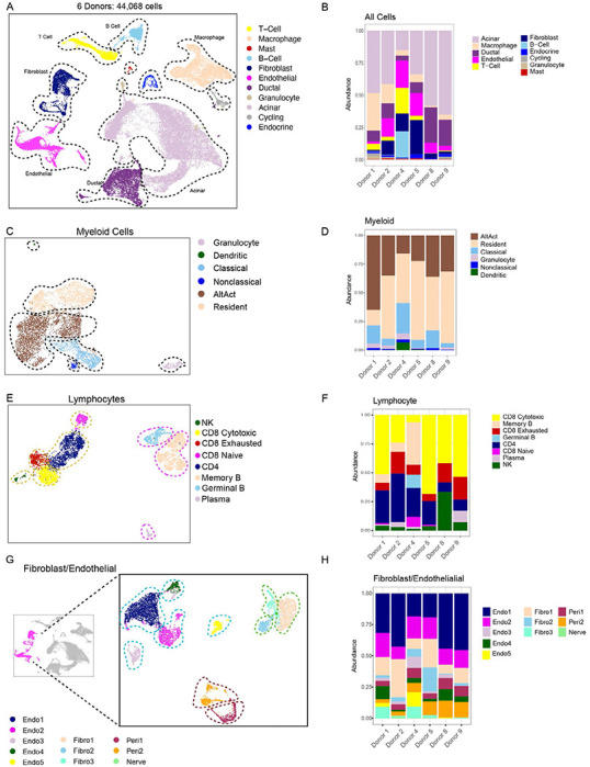
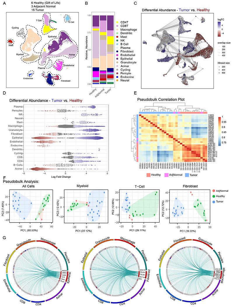
The adult healthy human pancreas has been poorly studied given lack of indication to obtain tissue from the pancreas in the absence of disease and rapid postmortem degradation. We obtained pancreata from brain dead donors thus avoiding any warm ischemia time. The 30 donors were diverse in age and race and had no known pancreas disease. Histopathological analysis of the samples revealed PanIN lesions in most individuals irrespective of age. Using a combination of multiplex immunohistochemistry, single cell RNA sequencing, and spatial transcriptomics, we provide the first ever characterization of the unique microenvironment of the adult human pancreas and of sporadic PanIN lesions. We compared healthy pancreata to pancreatic cancer and peritumoral tissue and observed distinct transcriptomic signatures in fibroblasts, and, to a lesser extent, macrophages. PanIN epithelial cells from healthy pancreata were remarkably transcriptionally similar to cancer cells, suggesting that neoplastic pathways are initiated early in tumorigenesis.
The causes underlying the onset of pancreatic cancer remain largely unknown, hampering early detection and prevention strategies. Here, we show that PanIN are abundant in healthy individuals and present at a much higher rate than the incidence of pancreatic cancer, setting the stage for efforts to elucidate the microenvironmental and cell intrinsic factors that restrain, or, conversely, promote, malignant progression.
鉴于在无疾病情况下获取胰腺组织缺乏指征以及死后胰腺迅速降解,对健康成人胰腺的研究较少。我们从脑死亡供体获取胰腺,从而避免了任何热缺血时间。30名供体年龄和种族各异,且无已知胰腺疾病。对样本的组织病理学分析显示,大多数个体均存在胰腺上皮内瘤变(PanIN)病变,与年龄无关。通过多重免疫组化、单细胞RNA测序和空间转录组学相结合的方法,我们首次对成人胰腺的独特微环境以及散发性PanIN病变进行了表征。我们将健康胰腺与胰腺癌及瘤周组织进行比较,观察到成纤维细胞以及程度较轻的巨噬细胞中存在不同的转录组特征。健康胰腺中的PanIN上皮细胞在转录水平上与癌细胞显著相似,这表明肿瘤发生途径在肿瘤发生早期就已启动。
胰腺癌发病的潜在原因在很大程度上仍不清楚,这阻碍了早期检测和预防策略的发展。在此,我们表明PanIN在健康个体中很常见,其出现率远高于胰腺癌的发病率,为阐明抑制或相反促进恶性进展的微环境和细胞内在因素的研究奠定了基础。