Isbister Julia C, Gray Belinda, Offen Sophie, Yeates Laura, Naoum Chris, Medi Caroline, Raju Hariharan, Semsarian Christopher, Puranik Rajesh, Sy Raymond W
Agnes Ginges Centre for Molecular Cardiology at Centenary Institute, University of Sydney, Sydney, New South Wales, Australia.
Faculty of Medicine and Heath, University of Sydney, Sydney, New South Wales, Australia.
Heart Rhythm O2. 2022 Oct 18;4(1):34-41. doi: 10.1016/j.hroo.2022.10.004. eCollection 2023 Jan.
Despite historically being considered a channelopathy, subtle structural changes have been reported in Brugada syndrome (BrS) on histopathology and cardiac magnetic resonance (CMR) imaging. It is not known if these structural changes progress over time.
The study sought to assess if structural changes in BrS evolve over time with serial CMR assessment and to investigate the utility of parametric mapping techniques to identify diffuse fibrosis in BrS.
Patients with a diagnosis of BrS based on international guidelines and normal CMR at least 3 years prior to the study period were invited to undergo repeat CMR. CMR images were analyzed de novo and compared at baseline and follow-up.
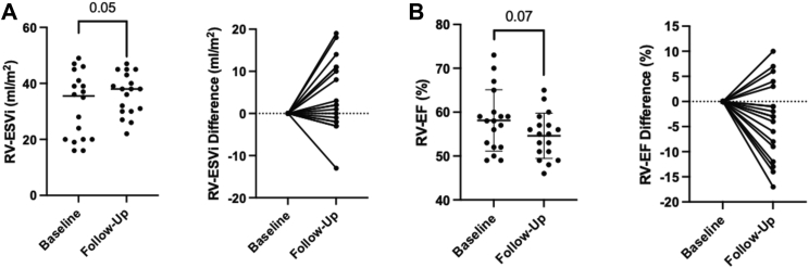
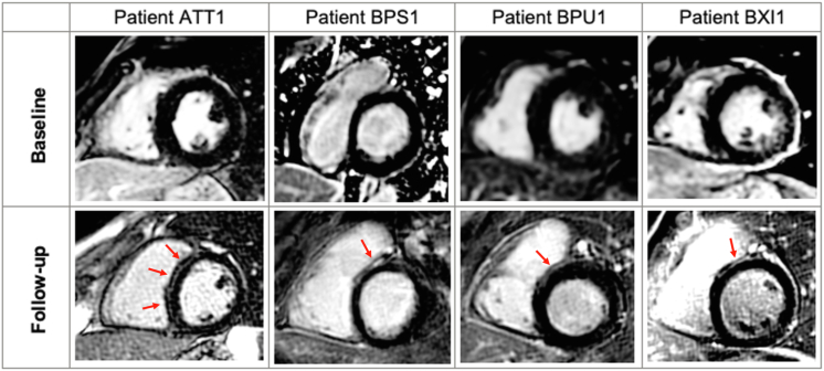
Eighteen patients with BrS (72% men; mean age at follow-up 47.4 ± 8.9 years) underwent serial CMR with an average of 5.0 ± 1.7 years between scans. No patients had late gadolinium enhancement (LGE) on baseline CMR, but 4 (22%) developed LGE on follow-up, typically localized to the right ventricular (RV) side of the basal septum. RV end-systolic volume increased over time ( .04) and was associated with a trend toward reduction in RV ejection fraction ( .07). Four patients showed a reduction in RV ejection fraction >10%. There was no evidence of diffuse myocardial fibrosis observed on parametric mapping.
Structural changes may evolve over time with development of focal fibrosis, evidenced by LGE on CMR in a significant proportion of patients with BrS. These findings have implications for our understanding of the pathological substrate in BrS and the longitudinal evaluation of patients with BrS.
尽管 Brugada 综合征(BrS)在历史上一直被认为是一种离子通道病,但组织病理学和心脏磁共振成像(CMR)已报道其存在细微的结构变化。目前尚不清楚这些结构变化是否会随时间进展。
本研究旨在通过连续 CMR 评估来评估 BrS 的结构变化是否随时间演变,并探讨参数映射技术在识别 BrS 弥漫性纤维化中的作用。
邀请根据国际指南诊断为 BrS 且在研究期前至少 3 年 CMR 正常的患者接受重复 CMR 检查。对 CMR 图像进行重新分析,并在基线和随访时进行比较。
18 例 BrS 患者(72%为男性;随访时平均年龄 47.4±8.9 岁)接受了连续 CMR 检查,两次扫描之间的平均时间为 5.0±1.7 年。基线 CMR 检查时无患者出现延迟钆增强(LGE),但随访时有 4 例(22%)出现 LGE,通常位于基底间隔的右心室(RV)侧。RV 收缩末期容积随时间增加(P=0.04),并与 RV 射血分数降低趋势相关(P=0.07)。4 例患者的 RV 射血分数降低>10%。参数映射未观察到弥漫性心肌纤维化的证据。
结构变化可能随时间演变,出现局灶性纤维化,在相当比例的 BrS 患者中,CMR 上的 LGE 可证明这一点。这些发现对我们理解 BrS 的病理基础以及 BrS 患者的纵向评估具有重要意义。