• 文献检索
  • 文档翻译
  • 深度研究
  • 学术资讯
  • Suppr Zotero 插件Zotero 插件
  • 邀请有礼
  • 套餐&价格
  • 历史记录
应用&插件
Suppr Zotero 插件Zotero 插件浏览器插件Mac 客户端Windows 客户端微信小程序
定价
高级版会员购买积分包购买API积分包
服务
文献检索文档翻译深度研究API 文档MCP 服务
关于我们
关于 Suppr公司介绍联系我们用户协议隐私条款
关注我们

Suppr 超能文献

核心技术专利:CN118964589B侵权必究
粤ICP备2023148730 号-1Suppr @ 2026

文献检索

告别复杂PubMed语法,用中文像聊天一样搜索,搜遍4000万医学文献。AI智能推荐,让科研检索更轻松。

立即免费搜索

文件翻译

保留排版,准确专业,支持PDF/Word/PPT等文件格式,支持 12+语言互译。

免费翻译文档

深度研究

AI帮你快速写综述,25分钟生成高质量综述,智能提取关键信息,辅助科研写作。

立即免费体验

基于深度学习的合成骨抑制模型在胸部 X 线片中肺结节检测的开发与验证。

Development and Validation of a Deep Learning-Based Synthetic Bone-Suppressed Model for Pulmonary Nodule Detection in Chest Radiographs.

机构信息

Department of Radiology and Research Institute of Radiological Science and Center for Clinical Image Data Science, Severance Hospital, Yonsei University College of Medicine, Seoul, Korea.

Department of Radiology, Dankook University Hospital, Cheonan, Chungnam Province, Republic of Korea.

出版信息

JAMA Netw Open. 2023 Jan 3;6(1):e2253820. doi: 10.1001/jamanetworkopen.2022.53820.

DOI:10.1001/jamanetworkopen.2022.53820
PMID:36719681
原文链接:https://pmc.ncbi.nlm.nih.gov/articles/PMC9890286/
Abstract

IMPORTANCE

Dual-energy chest radiography exhibits better sensitivity than single-energy chest radiography, partly due to its ability to remove overlying anatomical structures.

OBJECTIVES

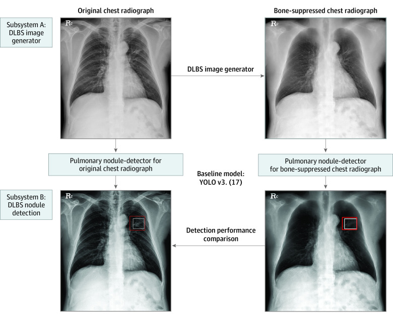
To develop and validate a deep learning-based synthetic bone-suppressed (DLBS) nodule-detection algorithm for pulmonary nodule detection on chest radiographs.

DESIGN, SETTING, AND PARTICIPANTS: This decision analytical modeling study used data from 3 centers between November 2015 and July 2019 from 1449 patients. The DLBS nodule-detection algorithm was trained using single-center data (institute 1) of 998 chest radiographs. The DLBS algorithm was validated using 2 external data sets (institute 2, 246 patients; and institute 3, 205 patients). Statistical analysis was performed from March to December 2021.

EXPOSURES

DLBS nodule-detection algorithm.

MAIN OUTCOMES AND MEASURES

The nodule-detection performance of DLBS model was compared with the convolution neural network nodule-detection algorithm (original model). Reader performance testing was conducted by 3 thoracic radiologists assisted by the DLBS algorithm or not. Sensitivity and false-positive markings per image (FPPI) were compared.

RESULTS

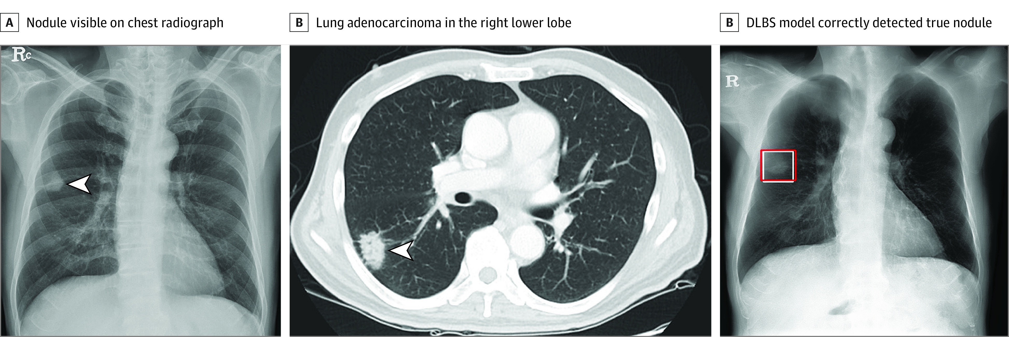
Training data consisted of 998 patients (539 men [54.0%]; mean [SD] age, 54.2 [9.82] years), and 2 external validation data sets consisted of 246 patients (133 men [54.1%]; mean [SD] age, 55.3 [8.7] years) and 205 patients (105 men [51.2%]; mean [SD] age, 51.8 [9.1] years). Using the external validation data set of institute 2, the bone-suppressed model showed higher sensitivity compared with that of the original model for nodule detection (91.5% [109 of 119] vs 79.8% [95 of 119]; P < .001). The overall mean of FPPI with the bone-suppressed model was reduced compared with the original model (0.07 [17 of 246] vs 0.09 [23 of 246]; P < .001). For the observer performance testing with the data of institute 3, the mean sensitivity of 3 radiologists was 77.5% (95% [CI], 69.9%-85.2%), whereas that of radiologists assisted by DLBS modeling was 92.1% (95% CI, 86.3%-97.3%; P < .001). The 3 radiologists had a reduced number of FPPI when assisted by the DLBS model (0.071 [95% CI, 0.041-0.111] vs 0.151 [95% CI, 0.111-0.210]; P < .001).

CONCLUSIONS AND RELEVANCE

This decision analytical modeling study found that the DLBS model was more sensitive to detecting pulmonary nodules on chest radiographs compared with the original model. These findings suggest that the DLBS model could be beneficial to radiologists in the detection of lung nodules in chest radiographs without need of the specialized equipment or increase of radiation dose.

摘要

重要性

双能胸部 X 射线摄影比单能胸部 X 射线摄影具有更好的敏感性,部分原因是它能够去除重叠的解剖结构。

目的

开发和验证一种基于深度学习的合成骨抑制(DLBS)结节检测算法,用于胸部 X 射线摄影中的肺结节检测。

设计、设置和参与者:这项决策分析模型研究使用了 2015 年 11 月至 2019 年 7 月期间来自 3 个中心的 1449 名患者的数据。DLBS 结节检测算法是使用单中心数据(机构 1)中的 998 张胸部 X 射线片进行训练的。DLBS 算法使用 2 个外部数据集(机构 2,246 名患者;机构 3,205 名患者)进行验证。统计分析于 2021 年 3 月至 12 月进行。

暴露

DLBS 结节检测算法。

主要结果和措施

比较了 DLBS 模型与卷积神经网络结节检测算法(原始模型)的结节检测性能。通过 3 名胸部放射科医生进行读者性能测试,协助或不协助 DLBS 算法。比较了敏感性和每张图像的假阳性标记数(FPPI)。

结果

训练数据包括 998 名患者(539 名男性[54.0%];平均[标准差]年龄,54.2[9.82]岁),2 个外部验证数据集包括 246 名患者(133 名男性[54.1%];平均[标准差]年龄,55.3[8.7]岁)和 205 名患者(105 名男性[51.2%];平均[标准差]年龄,51.8[9.1]岁)。使用机构 2 的外部验证数据集,与原始模型相比,骨抑制模型在结节检测方面具有更高的敏感性(91.5%[119 例中的 109 例]vs 79.8%[119 例中的 95 例];P < .001)。与原始模型相比,整体平均 FPPI 降低(0.07[246 例中的 17 例]vs 0.09[246 例中的 23 例];P < .001)。对于使用机构 3 数据的观察者性能测试,3 名放射科医生的平均敏感性为 77.5%(95%[置信区间],69.9%-85.2%),而在 DLBS 建模协助下,敏感性为 92.1%(95%CI,86.3%-97.3%;P < .001)。当使用 DLBS 模型时,3 名放射科医生的 FPPI 数量减少(0.071[95%CI,0.041-0.111]vs 0.151[95%CI,0.111-0.210];P < .001)。

结论和相关性

这项决策分析模型研究发现,与原始模型相比,DLBS 模型对胸部 X 射线摄影中的肺结节检测更敏感。这些发现表明,DLBS 模型可能有助于放射科医生在不使用专门设备或增加辐射剂量的情况下检测胸部 X 射线中的肺结节。

https://cdn.ncbi.nlm.nih.gov/pmc/blobs/37b8/9890286/b9c85ca7c1cf/jamanetwopen-e2253820-g002.jpg
https://cdn.ncbi.nlm.nih.gov/pmc/blobs/37b8/9890286/1001887395f0/jamanetwopen-e2253820-g001.jpg
https://cdn.ncbi.nlm.nih.gov/pmc/blobs/37b8/9890286/b9c85ca7c1cf/jamanetwopen-e2253820-g002.jpg
https://cdn.ncbi.nlm.nih.gov/pmc/blobs/37b8/9890286/1001887395f0/jamanetwopen-e2253820-g001.jpg
https://cdn.ncbi.nlm.nih.gov/pmc/blobs/37b8/9890286/b9c85ca7c1cf/jamanetwopen-e2253820-g002.jpg

相似文献

1
Development and Validation of a Deep Learning-Based Synthetic Bone-Suppressed Model for Pulmonary Nodule Detection in Chest Radiographs.基于深度学习的合成骨抑制模型在胸部 X 线片中肺结节检测的开发与验证。
JAMA Netw Open. 2023 Jan 3;6(1):e2253820. doi: 10.1001/jamanetworkopen.2022.53820.
2
Validation of a Deep Learning Algorithm for the Detection of Malignant Pulmonary Nodules in Chest Radiographs.深度学习算法在胸部 X 光片中检测恶性肺结节的验证。
JAMA Netw Open. 2020 Sep 1;3(9):e2017135. doi: 10.1001/jamanetworkopen.2020.17135.
3
Development and Validation of Deep Learning-based Automatic Detection Algorithm for Malignant Pulmonary Nodules on Chest Radiographs.基于深度学习的胸部 X 线片恶性肺结节自动检测算法的开发与验证。
Radiology. 2019 Jan;290(1):218-228. doi: 10.1148/radiol.2018180237. Epub 2018 Sep 25.
4
Development and Validation of a Deep Learning-Based Automated Detection Algorithm for Major Thoracic Diseases on Chest Radiographs.基于深度学习的胸部 X 线片主要胸部疾病自动检测算法的开发与验证。
JAMA Netw Open. 2019 Mar 1;2(3):e191095. doi: 10.1001/jamanetworkopen.2019.1095.
5
Deep Convolutional Neural Network-based Software Improves Radiologist Detection of Malignant Lung Nodules on Chest Radiographs.基于深度卷积神经网络的软件提高放射科医生在胸部 X 光片上检测恶性肺结节的能力。
Radiology. 2020 Jan;294(1):199-209. doi: 10.1148/radiol.2019182465. Epub 2019 Nov 12.
6
Optimal matrix size of chest radiographs for computer-aided detection on lung nodule or mass with deep learning.深度学习辅助肺结节或肿块计算机辅助检测的胸部 X 光片最佳矩阵大小。
Eur Radiol. 2020 Sep;30(9):4943-4951. doi: 10.1007/s00330-020-06892-9. Epub 2020 Apr 29.
7
AI-based improvement in lung cancer detection on chest radiographs: results of a multi-reader study in NLST dataset.基于人工智能的胸部 X 光肺癌检测改进:NLST 数据集的多读者研究结果。
Eur Radiol. 2021 Dec;31(12):9664-9674. doi: 10.1007/s00330-021-08074-7. Epub 2021 Jun 4.
8
Deep learning for chest radiograph diagnosis: A retrospective comparison of the CheXNeXt algorithm to practicing radiologists.深度学习在胸片诊断中的应用:CheXNeXt 算法与临床放射科医生的回顾性比较。
PLoS Med. 2018 Nov 20;15(11):e1002686. doi: 10.1371/journal.pmed.1002686. eCollection 2018 Nov.
9
Better performance of deep learning pulmonary nodule detection using chest radiography with pixel level labels in reference to computed tomography: data quality matters.使用带有像素级标签的胸部 X 光片对深度学习肺结节检测的性能提升:数据质量很重要。
Sci Rep. 2024 Jul 10;14(1):15967. doi: 10.1038/s41598-024-66530-y.
10
An Artificial Intelligence-Based Chest X-ray Model on Human Nodule Detection Accuracy From a Multicenter Study.基于人工智能的多中心研究中用于检测人类结节的胸部 X 射线模型。
JAMA Netw Open. 2021 Dec 1;4(12):e2141096. doi: 10.1001/jamanetworkopen.2021.41096.

引用本文的文献

1
A Systematic Review: The Role of Artificial Intelligence in Lung Cancer Screening in Detecting Lung Nodules on Chest X-Rays.一项系统评价:人工智能在肺癌筛查中对胸部X线片上肺结节检测的作用
Diagnostics (Basel). 2025 Jan 22;15(3):246. doi: 10.3390/diagnostics15030246.
2
Independent evaluation of the accuracy of 5 artificial intelligence software for detecting lung nodules on chest X-rays.对5种用于在胸部X光片上检测肺结节的人工智能软件准确性的独立评估。
Quant Imaging Med Surg. 2024 Aug 1;14(8):5288-5303. doi: 10.21037/qims-24-160. Epub 2024 Jul 25.
3
Assessment of Advanced Diagnostic Bronchoscopy Outcomes for Peripheral Lung Lesions: A Delphi Consensus Definition of Diagnostic Yield and Recommendations for Patient-centered Study Designs. An Official American Thoracic Society/American College of Chest Physicians Research Statement.

本文引用的文献

1
Bone Suppression on Chest Radiographs for Pulmonary Nodule Detection: Comparison between a Generative Adversarial Network and Dual-Energy Subtraction.胸部 X 线摄影检测肺结节时的骨抑制:生成对抗网络与双能减影的比较。
Korean J Radiol. 2022 Jan;23(1):139-149. doi: 10.3348/kjr.2021.0146.
2
Validation of a Deep Learning Algorithm for the Detection of Malignant Pulmonary Nodules in Chest Radiographs.深度学习算法在胸部 X 光片中检测恶性肺结节的验证。
JAMA Netw Open. 2020 Sep 1;3(9):e2017135. doi: 10.1001/jamanetworkopen.2020.17135.
3
Chest Radiograph Interpretation with Deep Learning Models: Assessment with Radiologist-adjudicated Reference Standards and Population-adjusted Evaluation.
评估外周肺病变高级诊断性支气管镜检查的结果:诊断产量的德尔福共识定义和以患者为中心的研究设计建议。美国胸科学会/美国胸科学会研究声明的官方声明。
Am J Respir Crit Care Med. 2024 Mar 15;209(6):634-646. doi: 10.1164/rccm.202401-0192ST.
4
Systematic review of deep learning image analyses for the diagnosis and monitoring of skin disease.深度学习图像分析用于皮肤病诊断和监测的系统评价
NPJ Digit Med. 2023 Sep 27;6(1):180. doi: 10.1038/s41746-023-00914-8.
5
New trend in artificial intelligence-based assistive technology for thoracic imaging.基于人工智能的胸像辅助技术的新趋势。
Radiol Med. 2023 Oct;128(10):1236-1249. doi: 10.1007/s11547-023-01691-w. Epub 2023 Aug 28.
深度学习模型在胸部 X 线片解读中的应用:使用经过放射科医师裁定的参考标准和人群校正评估进行评估。
Radiology. 2020 Feb;294(2):421-431. doi: 10.1148/radiol.2019191293. Epub 2019 Dec 3.
4
Deep Convolutional Neural Network-based Software Improves Radiologist Detection of Malignant Lung Nodules on Chest Radiographs.基于深度卷积神经网络的软件提高放射科医生在胸部 X 光片上检测恶性肺结节的能力。
Radiology. 2020 Jan;294(1):199-209. doi: 10.1148/radiol.2019182465. Epub 2019 Nov 12.
5
Identifying pulmonary nodules or masses on chest radiography using deep learning: external validation and strategies to improve clinical practice.利用深度学习技术在胸部 X 光片上识别肺结节或肿块:外部验证及改善临床实践的策略。
Clin Radiol. 2020 Jan;75(1):38-45. doi: 10.1016/j.crad.2019.08.005. Epub 2019 Sep 11.
6
Development of a deep neural network for generating synthetic dual-energy chest x-ray images with single x-ray exposure.基于单次 X 射线曝光生成合成双能胸部 X 射线图像的深度神经网络开发。
Phys Med Biol. 2019 May 31;64(11):115017. doi: 10.1088/1361-6560/ab1cee.
7
Separation of bones from soft tissue in chest radiographs: Anatomy-specific orientation-frequency-specific deep neural network convolution.胸部 X 光片中骨与软组织的分离:解剖特定位向-频率特定深度神经网络卷积。
Med Phys. 2019 May;46(5):2232-2242. doi: 10.1002/mp.13468. Epub 2019 Mar 28.
8
Deep learning for chest radiograph diagnosis: A retrospective comparison of the CheXNeXt algorithm to practicing radiologists.深度学习在胸片诊断中的应用:CheXNeXt 算法与临床放射科医生的回顾性比较。
PLoS Med. 2018 Nov 20;15(11):e1002686. doi: 10.1371/journal.pmed.1002686. eCollection 2018 Nov.
9
Development and Validation of Deep Learning-based Automatic Detection Algorithm for Malignant Pulmonary Nodules on Chest Radiographs.基于深度学习的胸部 X 线片恶性肺结节自动检测算法的开发与验证。
Radiology. 2019 Jan;290(1):218-228. doi: 10.1148/radiol.2018180237. Epub 2018 Sep 25.
10
Training and Validating a Deep Convolutional Neural Network for Computer-Aided Detection and Classification of Abnormalities on Frontal Chest Radiographs.训练和验证用于胸部正位X光片异常的计算机辅助检测与分类的深度卷积神经网络
Invest Radiol. 2017 May;52(5):281-287. doi: 10.1097/RLI.0000000000000341.