Department of Physics and Astronomy, University of British Columbia, Vancouver, British Columbia, Canada.
Department of Pediatrics, University of British Columbia, Vancouver, British Columbia, Canada.
Brain Pathol. 2023 Nov;33(6):e13150. doi: 10.1111/bpa.13150. Epub 2023 Jan 31.
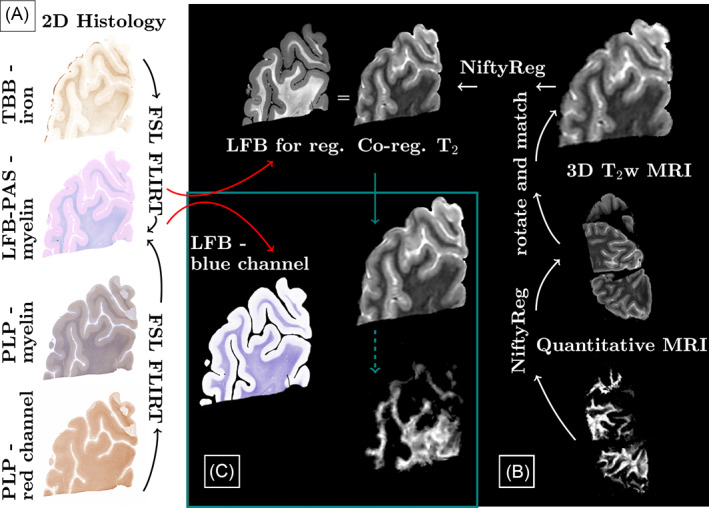
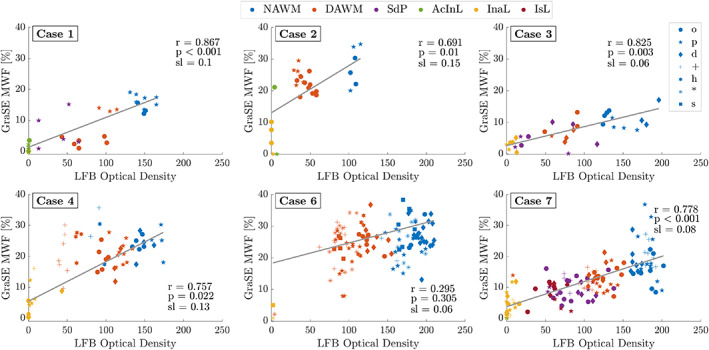
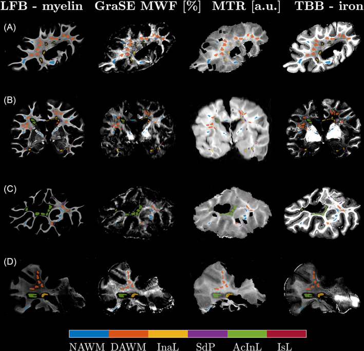
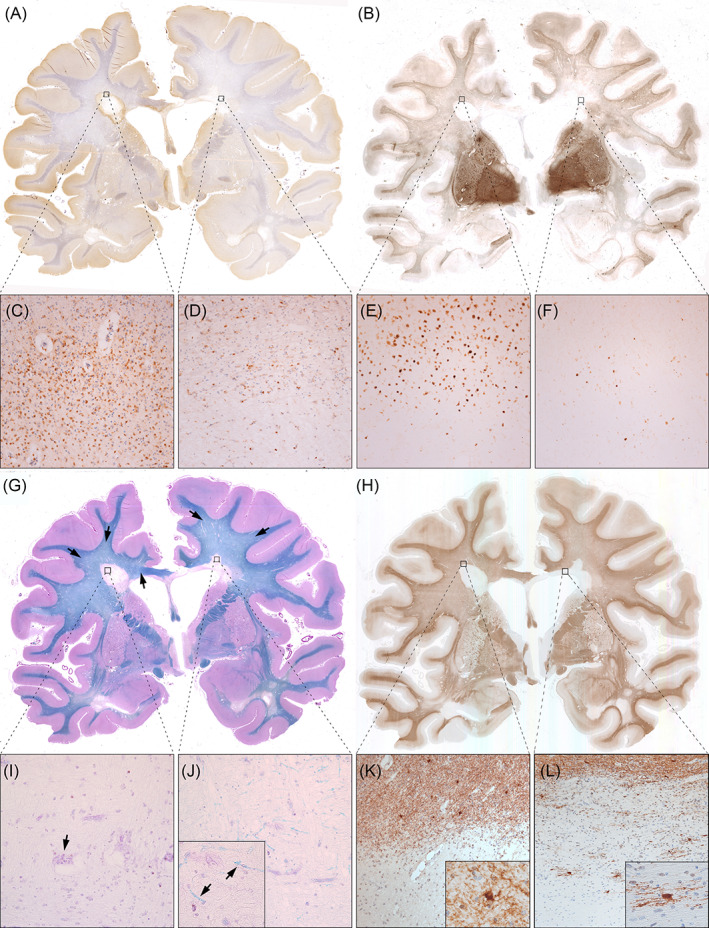
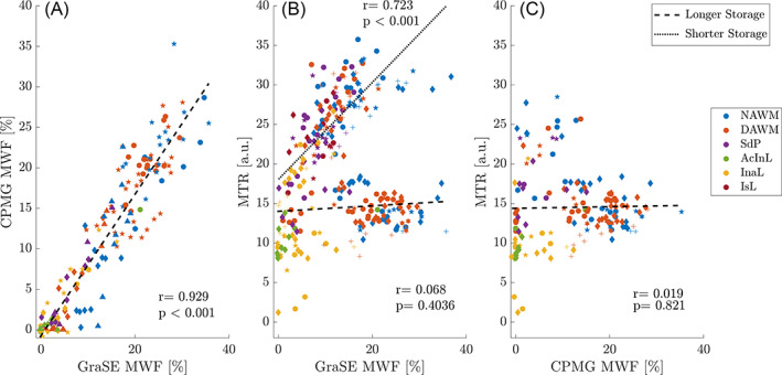
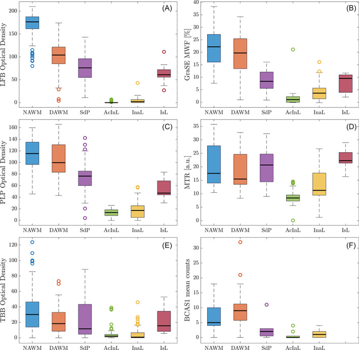
Magnetic resonance imaging (MRI) of focal or diffuse myelin damage or remyelination may provide important insights into disease progression and potential treatment efficacy in multiple sclerosis (MS). We performed post-mortem MRI and histopathological myelin measurements in seven progressive MS cases to evaluate the ability of three myelin-sensitive MRI scans to distinguish different stages of MS pathology, particularly chronic demyelinated and remyelinated lesions. At 3 Tesla, we acquired two different myelin water imaging (MWI) scans and magnetisation transfer ratio (MTR) data. Histopathology included histochemical stainings for myelin phospholipids (LFB) and iron as well as immunohistochemistry for myelin proteolipid protein (PLP), CD68 (phagocytosing microglia/macrophages) and BCAS1 (remyelinating oligodendrocytes). Mixed-effects modelling determined which histopathological metric best predicted MWF and MTR in normal-appearing and diffusely abnormal white matter, active/inactive, inactive, remyelinated and ischemic lesions. Both MWI measures correlated well with each other and histology across regions, reflecting the different stages of MS pathology. MTR data showed a considerable influence of components other than myelin and a strong dependency on tissue storage duration. Both MRI and histology revealed increased myelin densities in inactive compared with active/inactive lesions. Chronic inactive lesions harboured single scattered myelin fibres indicative of low-level remyelination. Mixed-effects modelling showed that smaller differences between white matter areas were linked to PLP densities and only to a small extent confounded by iron. MWI reflects differences in myelin lipids and proteins across various levels of myelin densities encountered in MS, including low-level remyelination in chronic inactive lesions.
磁共振成像(MRI)对局灶性或弥漫性髓鞘损伤或髓鞘再生的研究,可能为多发性硬化症(MS)的疾病进展和潜在治疗效果提供重要的见解。我们对 7 例进行性 MS 病例进行了死后 MRI 和组织病理学髓鞘测量,以评估三种敏感髓鞘 MRI 扫描区分 MS 病理学不同阶段(尤其是慢性脱髓鞘和髓鞘再生病变)的能力。在 3T 下,我们获得了两种不同的髓鞘水成像(MWI)扫描和磁化传递率(MTR)数据。组织病理学包括髓磷脂磷脂(LFB)和铁的组织化学染色以及髓鞘蛋白脂蛋白(PLP)、CD68(吞噬性小胶质细胞/巨噬细胞)和 BCAS1(髓鞘再生少突胶质细胞)的免疫组化染色。混合效应模型确定了哪些组织病理学指标最能预测正常表现和弥漫性异常白质、活动/非活动、非活动、髓鞘再生和缺血性病变中的 MWF 和 MTR。两种 MWI 测量方法与彼此以及整个区域的组织学均具有良好的相关性,反映了 MS 病理学的不同阶段。MTR 数据显示,除了髓鞘之外,其他成分对其有很大影响,并且强烈依赖于组织储存时间。MRI 和组织学均显示,与活动/非活动病变相比,非活动病变中的髓鞘密度增加。慢性非活动病变中存在单个散在的髓鞘纤维,提示低水平的髓鞘再生。混合效应模型表明,白质区域之间较小的差异与 PLP 密度有关,仅在很小程度上受到铁的混杂。MWI 反映了 MS 中各种髓鞘密度水平下髓鞘脂质和蛋白质的差异,包括慢性非活动病变中的低水平髓鞘再生。