Department of Clinical Infectious Diseases, Toyama University Graduate School of Medicine and Pharmaceutical Sciences, University of Toyama, 2630 Sugitani, Toyama, 930-0194, Japan.
Department of Emergency Medicine, Toyama University Graduate School of Medicine and Pharmaceutical Sciences, University of Toyama, 2630 Sugitani, Toyama, 930-0194, Japan.
Eur Radiol. 2023 Jul;33(7):4713-4722. doi: 10.1007/s00330-023-09427-0. Epub 2023 Feb 3.
To examine the radiological patterns specifically associated with hypoxemic respiratory failure in patients with coronavirus disease (COVID-19).
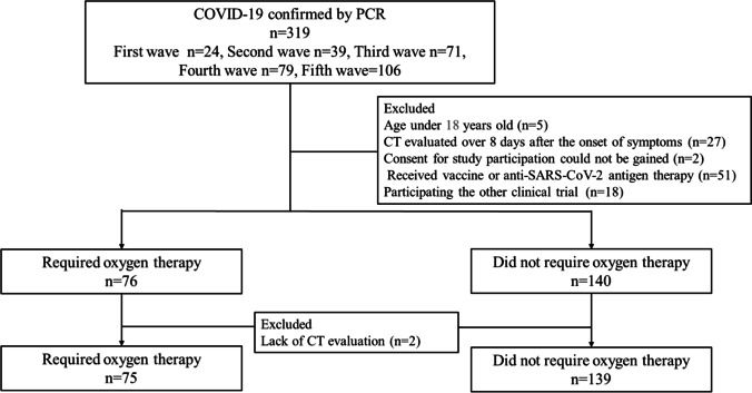
We enrolled patients with COVID-19 confirmed by qPCR in this prospective observational cohort study. We explored the association of clinical, radiological, and microbiological data with the development of hypoxemic respiratory failure after COVID-19 onset. Semi-quantitative CT scores and dominant CT patterns were retrospectively determined for each patient. The microbiological evaluation included checking the SARS-CoV-2 viral load by qPCR using nasal swab and serum specimens.
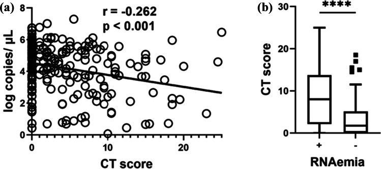
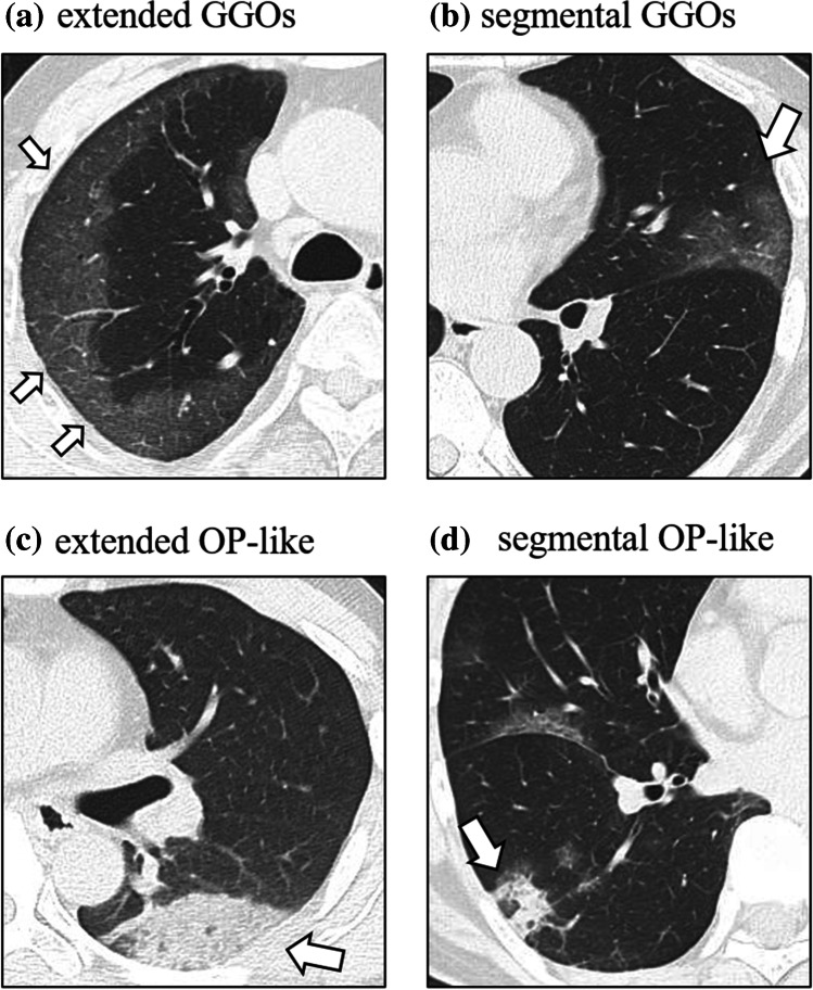
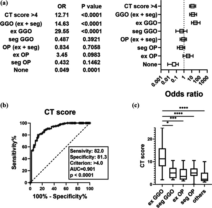
Of the 214 eligible patients, 75 developed hypoxemic respiratory failure and 139 did not. The CT score was significantly higher in patients who developed hypoxemic respiratory failure than in those did not (median [interquartile range]: 9 [6-14] vs 0 [0-3]; p < 0.001). The dominant CT patterns were subpleural ground-glass opacities (GGOs) extending beyond the segmental area (n = 44); defined as "extended GGOs." Multivariable analysis showed that hypoxemic respiratory failure was significantly associated with extended GGOs (odds ratio [OR] 29.6; 95% confidence interval [CI], 9.3-120; p < 0.001), and a CT score > 4 (OR 12.7; 95% CI, 5.3-33; p < 0.001). The incidence of RNAemia was significantly higher in patients with extended GGOs (58.3%) than in those without any pulmonary lesion (14.7%; p < 0.001).
Extended GGOs along the subpleural area were strongly associated with hypoxemia and viremia in patients with COVID-19.
• Extended ground-glass opacities (GGOs) along the subpleural area and a CT score > 4, in the early phase of COVID-19, were independently associated with the development of hypoxemic respiratory failure. • The absence of pulmonary lesions on CT in the early phase of COVID-19 was associated with a lower risk of developing hypoxemic respiratory failure. • Compared to patients with other CT findings, the extended GGOs and a higher CT score were also associated with a higher incidence of RNAemia.
研究与 COVID-19 患者低氧性呼吸衰竭相关的特定放射学模式。
我们对这项前瞻性观察队列研究中通过 qPCR 确诊的 COVID-19 患者进行了研究。我们探讨了临床、放射学和微生物学数据与 COVID-19 发病后低氧性呼吸衰竭的发生之间的关系。为每位患者回顾性确定半定量 CT 评分和主要 CT 模式。微生物学评估包括使用鼻拭子和血清样本通过 qPCR 检查 SARS-CoV-2 病毒载量。
在 214 名合格患者中,75 名患者发生低氧性呼吸衰竭,139 名患者未发生。与未发生低氧性呼吸衰竭的患者相比,发生低氧性呼吸衰竭的患者 CT 评分明显更高(中位数[四分位数范围]:9 [6-14] vs 0 [0-3];p<0.001)。主要 CT 模式为胸膜下磨玻璃影(GGO)延伸超出节段区域(n=44);定义为“扩展 GGO”。多变量分析显示,低氧性呼吸衰竭与扩展 GGO 显著相关(优势比[OR] 29.6;95%置信区间[CI],9.3-120;p<0.001),且 CT 评分>4(OR 12.7;95% CI,5.3-33;p<0.001)。扩展 GGO 患者的 RNA 血症发生率明显高于无任何肺部病变的患者(58.3% vs 14.7%;p<0.001)。
COVID-19 患者胸膜下区域的扩展 GGO 与低氧血症和病毒血症密切相关。