Zhang Xiangmin, Song Wei, Liu Xingli, Lyu Liang
Department of Radiology, The First People's Hospital of Yunnan Province, No. 157 Jinbi Road, Kunming, 650032, Yunnan, China.
The Affiliated Hospital Kunming University of Science and Technology, Kunming, Yunnan, China.
Jpn J Radiol. 2020 May;38(5):407-408. doi: 10.1007/s11604-020-00945-1. Epub 2020 Mar 18.
Knowledge of CT characteristics of COVID-19 pneumonia might be helpful to the early diagnosis and treatment of patients, and to control the spread of infection.
The chest CT images of the patient were collected to describe the CT manifestations and characteristics, and they were compared with the previous studies.
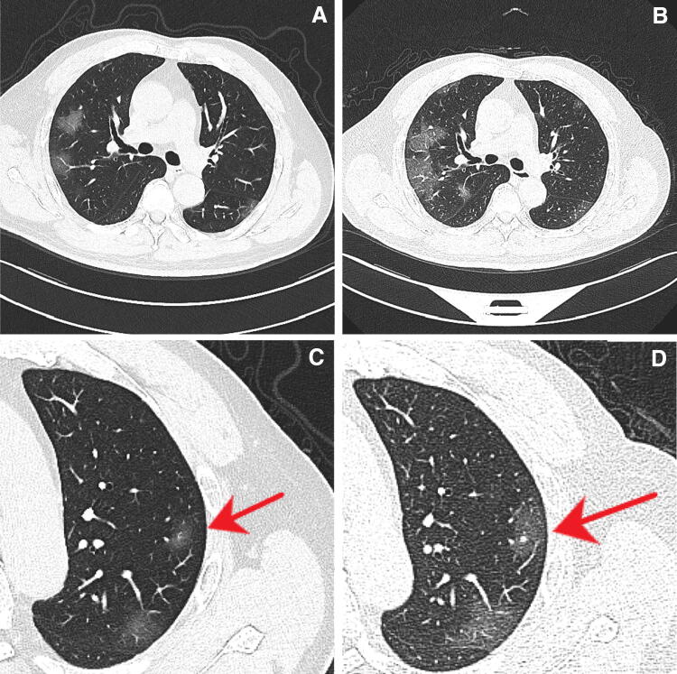
Multiple patchy ground-glass opacities (GGOs) were seen in bilateral lung, mostly in subpleural areas. They progressed within 3 days, and nodular GGOs were also seen together with subpleural patchy GGOs.
Our case of COVID-19 pneumonia showed multiple subpleural GGOs in bilateral lung, rapid progression, and it also accompanied nodular GGOs on chest CT. These findings were consistent with the previous reports, and they might be useful for early detection and evaluation of severity of COVID-19 pneumonia.
了解新型冠状病毒肺炎(COVID-19)的胸部CT特征可能有助于患者的早期诊断和治疗,并控制感染传播。
收集患者的胸部CT图像以描述CT表现和特征,并与先前的研究进行比较。
双侧肺可见多发斑片状磨玻璃影(GGO),主要位于胸膜下区域。它们在3天内进展,还可见结节状GGO与胸膜下斑片状GGO同时存在。
我们的COVID-19肺炎病例显示双侧肺多发胸膜下GGO,进展迅速,胸部CT还伴有结节状GGO。这些发现与先前的报道一致,可能有助于COVID-19肺炎的早期检测和严重程度评估。