• 文献检索
  • 文档翻译
  • 深度研究
  • 学术资讯
  • Suppr Zotero 插件Zotero 插件
  • 邀请有礼
  • 套餐&价格
  • 历史记录
应用&插件
Suppr Zotero 插件Zotero 插件浏览器插件Mac 客户端Windows 客户端微信小程序
定价
高级版会员购买积分包购买API积分包
服务
文献检索文档翻译深度研究API 文档MCP 服务
关于我们
关于 Suppr公司介绍联系我们用户协议隐私条款
关注我们

Suppr 超能文献

核心技术专利:CN118964589B侵权必究
粤ICP备2023148730 号-1Suppr @ 2026

文献检索

告别复杂PubMed语法,用中文像聊天一样搜索,搜遍4000万医学文献。AI智能推荐,让科研检索更轻松。

立即免费搜索

文件翻译

保留排版,准确专业,支持PDF/Word/PPT等文件格式,支持 12+语言互译。

免费翻译文档

深度研究

AI帮你快速写综述,25分钟生成高质量综述,智能提取关键信息,辅助科研写作。

立即免费体验

基于注意力的上下文感知深度框架用于婴儿脑皮质下分割。

An attention-based context-informed deep framework for infant brain subcortical segmentation.

机构信息

Department of Radiology and BRIC, University of North Carolina at Chapel Hill, Chapel Hill, NC 27599, USA.

Department of Radiology and BRIC, University of North Carolina at Chapel Hill, Chapel Hill, NC 27599, USA.

出版信息

Neuroimage. 2023 Apr 1;269:119931. doi: 10.1016/j.neuroimage.2023.119931. Epub 2023 Feb 4.

DOI:10.1016/j.neuroimage.2023.119931
PMID:36746299
原文链接:https://pmc.ncbi.nlm.nih.gov/articles/PMC10241225/
Abstract

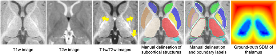
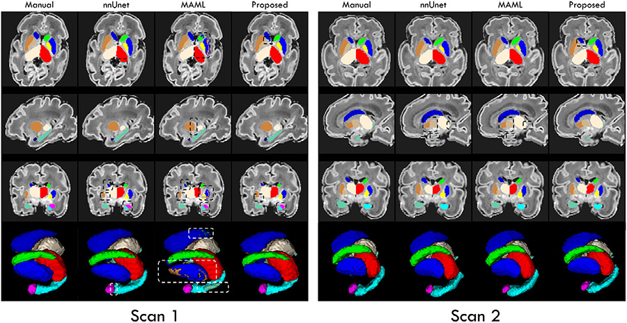
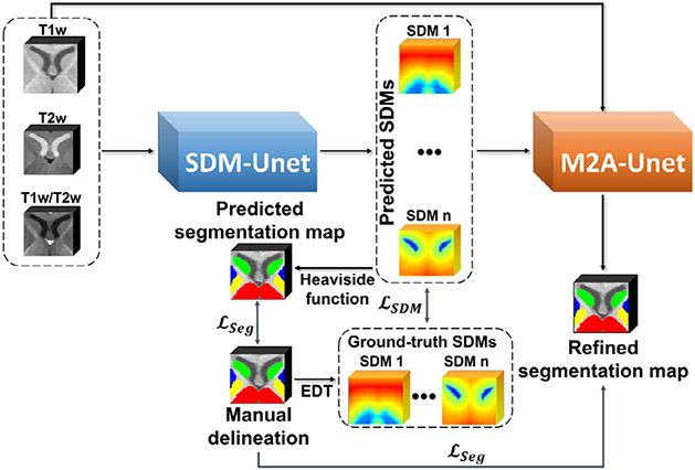
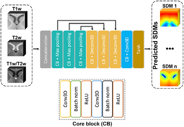
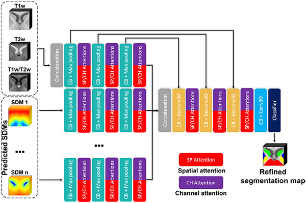
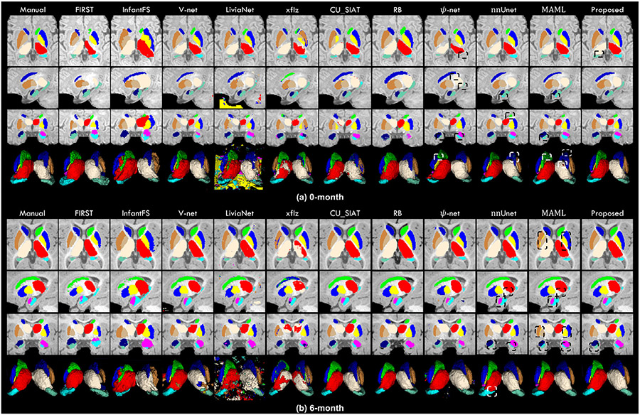
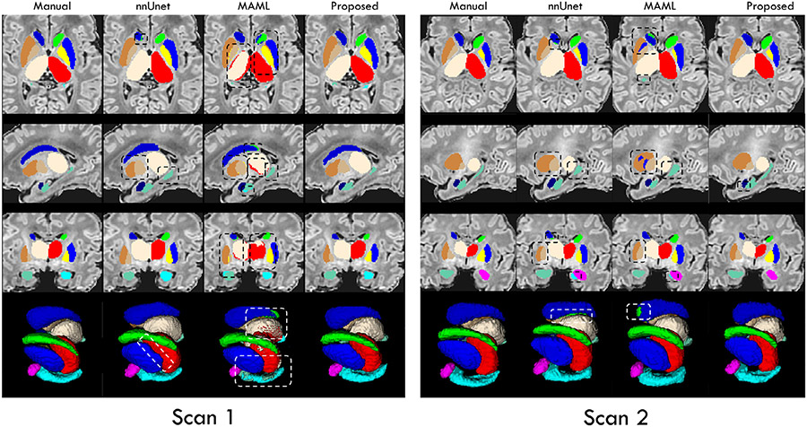
Precise segmentation of subcortical structures from infant brain magnetic resonance (MR) images plays an essential role in studying early subcortical structural and functional developmental patterns and diagnosis of related brain disorders. However, due to the dynamic appearance changes, low tissue contrast, and tiny subcortical size in infant brain MR images, infant subcortical segmentation is a challenging task. In this paper, we propose a context-guided, attention-based, coarse-to-fine deep framework to precisely segment the infant subcortical structures. At the coarse stage, we aim to directly predict the signed distance maps (SDMs) from multi-modal intensity images, including T1w, T2w, and the ratio of T1w and T2w images, with an SDM-Unet, which can leverage the spatial context information, including the structural position information and the shape information of the target structure, to generate high-quality SDMs. At the fine stage, the predicted SDMs, which encode spatial-context information of each subcortical structure, are integrated with the multi-modal intensity images as the input to a multi-source and multi-path attention Unet (M2A-Unet) for achieving refined segmentation. Both the 3D spatial and channel attention blocks are added to guide the M2A-Unet to focus more on the important subregions and channels. We additionally incorporate the inner and outer subcortical boundaries as extra labels to help precisely estimate the ambiguous boundaries. We validate our method on an infant MR image dataset and on an unrelated neonatal MR image dataset. Compared to eleven state-of-the-art methods, the proposed framework consistently achieves higher segmentation accuracy in both qualitative and quantitative evaluations of infant MR images and also exhibits good generalizability in the neonatal dataset.

摘要

精确分割婴儿脑磁共振(MR)图像的皮质下结构对于研究早期皮质下结构和功能发育模式以及诊断相关脑疾病至关重要。然而,由于婴儿脑 MR 图像中的动态外观变化、低组织对比度和微小的皮质下尺寸,婴儿皮质下分割是一项具有挑战性的任务。在本文中,我们提出了一种基于上下文引导、注意力机制的粗到精深度框架,用于精确分割婴儿皮质下结构。在粗阶段,我们旨在直接从多模态强度图像(包括 T1w、T2w 和 T1w 与 T2w 图像的比值)预测符号距离图(SDM),使用 SDM-Unet 可以利用空间上下文信息,包括目标结构的结构位置信息和形状信息,生成高质量的 SDM。在精细阶段,预测的 SDM 编码了每个皮质下结构的空间上下文信息,与多模态强度图像一起作为输入,输入到多源多路径注意力 U 型网络(M2A-Unet)中,以实现精细分割。我们还添加了 3D 空间注意力块和通道注意力块,以引导 M2A-Unet 更加关注重要的子区域和通道。我们还将内外皮质下边界作为额外的标签,以帮助更准确地估计模糊边界。我们在婴儿 MR 图像数据集和不相关的新生儿 MR 图像数据集上验证了我们的方法。与十一种最先进的方法相比,所提出的框架在婴儿 MR 图像的定性和定量评估中始终实现了更高的分割精度,并且在新生儿数据集上也表现出良好的泛化能力。

https://cdn.ncbi.nlm.nih.gov/pmc/blobs/fe84/10241225/e42b11ddcea2/nihms-1901215-f0009.jpg
https://cdn.ncbi.nlm.nih.gov/pmc/blobs/fe84/10241225/004cb0c4c92c/nihms-1901215-f0001.jpg
https://cdn.ncbi.nlm.nih.gov/pmc/blobs/fe84/10241225/3d39bdb231db/nihms-1901215-f0002.jpg
https://cdn.ncbi.nlm.nih.gov/pmc/blobs/fe84/10241225/27f5c97c949a/nihms-1901215-f0003.jpg
https://cdn.ncbi.nlm.nih.gov/pmc/blobs/fe84/10241225/163dac3d4a6d/nihms-1901215-f0004.jpg
https://cdn.ncbi.nlm.nih.gov/pmc/blobs/fe84/10241225/2a51d79176d9/nihms-1901215-f0005.jpg
https://cdn.ncbi.nlm.nih.gov/pmc/blobs/fe84/10241225/5bee316f18df/nihms-1901215-f0006.jpg
https://cdn.ncbi.nlm.nih.gov/pmc/blobs/fe84/10241225/6610dc0ca008/nihms-1901215-f0007.jpg
https://cdn.ncbi.nlm.nih.gov/pmc/blobs/fe84/10241225/94f289966e07/nihms-1901215-f0008.jpg
https://cdn.ncbi.nlm.nih.gov/pmc/blobs/fe84/10241225/e42b11ddcea2/nihms-1901215-f0009.jpg

相似文献

1
An attention-based context-informed deep framework for infant brain subcortical segmentation.基于注意力的上下文感知深度框架用于婴儿脑皮质下分割。
Neuroimage. 2023 Apr 1;269:119931. doi: 10.1016/j.neuroimage.2023.119931. Epub 2023 Feb 4.
2
A Deep Spatial Context Guided Framework for Infant Brain Subcortical Segmentation.一种用于婴儿脑皮质下分割的深度空间上下文引导框架。
Med Image Comput Comput Assist Interv. 2020 Oct;12267:646-656. doi: 10.1007/978-3-030-59728-3_63. Epub 2020 Sep 29.
3
Anatomical Attention Guided Deep Networks for ROI Segmentation of Brain MR Images.基于解剖注意力引导的深度网络的脑 MRI 图像 ROI 分割。
IEEE Trans Med Imaging. 2020 Jun;39(6):2000-2012. doi: 10.1109/TMI.2019.2962792. Epub 2019 Dec 30.
4
A 3D hierarchical cross-modality interaction network using transformers and convolutions for brain glioma segmentation in MR images.一种使用变换和卷积的 3D 层次跨模态交互网络,用于磁共振图像中的脑胶质瘤分割。
Med Phys. 2024 Nov;51(11):8371-8389. doi: 10.1002/mp.17354. Epub 2024 Aug 13.
5
LINKS: learning-based multi-source IntegratioN frameworK for Segmentation of infant brain images.LINKS:用于婴儿脑图像分割的基于学习的多源集成框架
Neuroimage. 2015 Mar;108:160-72. doi: 10.1016/j.neuroimage.2014.12.042. Epub 2014 Dec 22.
6
A Robust and Accurate Deep-learning-based Method for the Segmentation of Subcortical Brain: Cross-dataset Evaluation of Generalization Performance.基于深度学习的稳健准确的皮质下脑分割方法:泛化性能的跨数据集评估。
Magn Reson Med Sci. 2021 Jun 1;20(2):166-174. doi: 10.2463/mrms.mp.2019-0199. Epub 2020 May 11.
7
Multi-Modal Brain Tumor Data Completion Based on Reconstruction Consistency Loss.基于重建一致性损失的多模态脑肿瘤数据补全。
J Digit Imaging. 2023 Aug;36(4):1794-1807. doi: 10.1007/s10278-022-00697-6. Epub 2023 Mar 1.
8
Coarse-to-fine prior-guided attention network for multi-structure segmentation on dental panoramic radiographs.基于粗到精先验引导注意力网络的口腔全景片多结构分割。
Phys Med Biol. 2023 Oct 26;68(21). doi: 10.1088/1361-6560/ad0218.
9
[Fully Automatic Glioma Segmentation Algorithm of Magnetic Resonance Imaging Based on 3D-UNet With More Global Contextual Feature Extraction: An Improvement on Insufficient Extraction of Global Features].基于具有更多全局上下文特征提取的3D-UNet的磁共振成像全自动胶质瘤分割算法:对全局特征提取不足的改进
Sichuan Da Xue Xue Bao Yi Xue Ban. 2024 Mar 20;55(2):447-454. doi: 10.12182/20240360208.
10
APRNet: A 3D Anisotropic Pyramidal Reversible Network With Multi-Modal Cross-Dimension Attention for Brain Tissue Segmentation in MR Images.APRNet:一种具有多模态跨维度注意力的 3D 各向异性金字塔可逆网络,用于磁共振图像中的脑组织分割。
IEEE J Biomed Health Inform. 2022 Feb;26(2):749-761. doi: 10.1109/JBHI.2021.3093932. Epub 2022 Feb 4.

引用本文的文献

1
Baby Open Brains: An open-source dataset of infant brain segmentations.婴儿开放脑图谱:一个婴儿脑部分割的开源数据集。
Sci Data. 2025 Aug 14;12(1):1423. doi: 10.1038/s41597-025-05404-y.
2
AirSeg: Learnable Interconnected Attention Framework for Robust Airway Segmentation.AirSeg:用于稳健气道分割的可学习互联注意力框架
J Imaging Inform Med. 2025 May 22. doi: 10.1007/s10278-025-01545-z.
3
Baby Open Brains: An Open-Source Repository of Infant Brain Segmentations.婴儿开放大脑:一个婴儿脑部分割的开源库。

本文引用的文献

1
A 4D infant brain volumetric atlas based on the UNC/UMN baby connectome project (BCP) cohort.基于 UNC/UMN 婴儿连接组计划 (BCP) 队列的 4D 婴儿脑容量图谱。
Neuroimage. 2022 Jun;253:119097. doi: 10.1016/j.neuroimage.2022.119097. Epub 2022 Mar 14.
2
Anatomically constrained squeeze-and-excitation graph attention network for cortical surface parcellation.用于皮质表面分割的解剖学约束挤压与激励图注意力网络
Comput Biol Med. 2022 Jan;140:105113. doi: 10.1016/j.compbiomed.2021.105113. Epub 2021 Dec 4.
3
Visualization of iron-rich subcortical structures in non-human primates in vivo by quantitative susceptibility mapping at 3T MRI.
bioRxiv. 2024 Oct 14:2024.10.02.616147. doi: 10.1101/2024.10.02.616147.
4
Multi-kernel Learning Fusion Algorithm Based on RNN and GRU for ASD Diagnosis and Pathogenic Brain Region Extraction.基于 RNN 和 GRU 的多内核学习融合算法在 ASD 诊断和致病脑区提取中的应用。
Interdiscip Sci. 2024 Sep;16(3):755-768. doi: 10.1007/s12539-024-00629-8. Epub 2024 Apr 29.
在 3T MRI 下通过定量磁化率映射对非人类灵长类动物体内富含铁的皮质下结构进行可视化。
Neuroimage. 2021 Nov 1;241:118429. doi: 10.1016/j.neuroimage.2021.118429. Epub 2021 Jul 24.
4
A Deep Spatial Context Guided Framework for Infant Brain Subcortical Segmentation.一种用于婴儿脑皮质下分割的深度空间上下文引导框架。
Med Image Comput Comput Assist Interv. 2020 Oct;12267:646-656. doi: 10.1007/978-3-030-59728-3_63. Epub 2020 Sep 29.
5
Multi-Site Infant Brain Segmentation Algorithms: The iSeg-2019 Challenge.多站点婴儿脑分割算法:iSeg-2019 挑战赛。
IEEE Trans Med Imaging. 2021 May;40(5):1363-1376. doi: 10.1109/TMI.2021.3055428. Epub 2021 Apr 30.
6
nnU-Net: a self-configuring method for deep learning-based biomedical image segmentation.nnU-Net:一种基于深度学习的生物医学图像分割的自配置方法。
Nat Methods. 2021 Feb;18(2):203-211. doi: 10.1038/s41592-020-01008-z. Epub 2020 Dec 7.
7
Infant FreeSurfer: An automated segmentation and surface extraction pipeline for T1-weighted neuroimaging data of infants 0-2 years.婴儿 FreeSurfer:用于 0-2 岁婴儿 T1 加权神经影像学数据的自动分割和表面提取管道。
Neuroimage. 2020 Sep;218:116946. doi: 10.1016/j.neuroimage.2020.116946. Epub 2020 May 20.
8
A Longitudinal MRI Study of Amygdala and Hippocampal Subfields for Infants with Risk of Autism.一项针对有自闭症风险婴儿的杏仁核和海马亚区的纵向磁共振成像研究。
Graph Learn Med Imaging (2019). 2019 Oct;11849:164-171. doi: 10.1007/978-3-030-35817-4_20. Epub 2019 Nov 14.
9
ψ-Net: Stacking Densely Convolutional LSTMs for Sub-Cortical Brain Structure Segmentation.ψ-Net:用于皮质下脑结构分割的堆叠密集卷积 LSTM。
IEEE Trans Med Imaging. 2020 Sep;39(9):2806-2817. doi: 10.1109/TMI.2020.2975642. Epub 2020 Feb 24.
10
Unpaired Multi-Modal Segmentation via Knowledge Distillation.基于知识蒸馏的非配对多模态分割。
IEEE Trans Med Imaging. 2020 Jul;39(7):2415-2425. doi: 10.1109/TMI.2019.2963882. Epub 2020 Feb 3.