Khani Vahid, Momeni Moghaddam Amin, Hatami Behzad
Department of Radiology, Taleghani Hospital, Shahid Beheshti University of Medical Sciences, Tehran, Iran.
Gastroenterology and Liver Diseases Research Center, Research Institute for Gastroenterology and Liver Diseases, Shahid Beheshti University of Medical Sciences, Tehran, Iran.
Gastroenterol Hepatol Bed Bench. 2022;15(4):360-365. doi: 10.22037/ghfbb.v15i4.2480.
This study aimed to evaluate hepatic steatosis index (HSI) as a non-invasive tool in diagnosing and predicting nonalcoholic fatty liver disease (NAFLD) and to compare it with abdominal ultrasound as the gold standard tool.
NAFLD is a general term attributed to the deposition of adipose tissue in the liver leading to hepatitis, fibrosis, cirrhosis, and also hepatocellular carcinoma (HCC). Rapid and valid screening can remarkably prevent the progression of this disease.
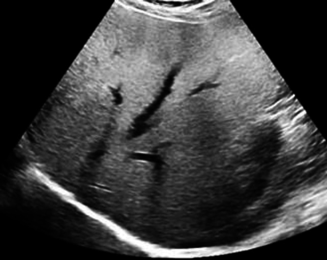
A total of 464 people were included in the present study based on inclusion criteria. Patients were evaluated for body mass index (BMI), AST, ALT, and ALP indices. The liver echogenicity of patients was evaluated by abdominal ultrasound technique.
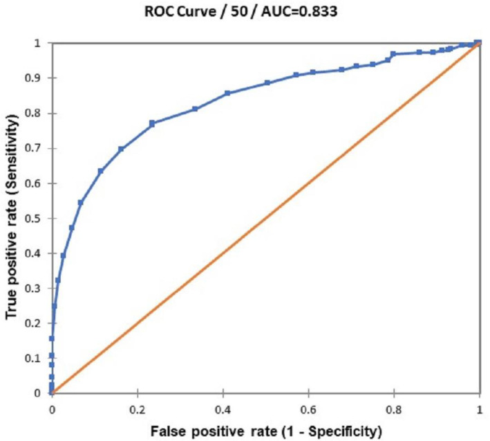
The results showed that out of 464 people included in the study, 32.33% represented fatty liver. It was found that 79.1% of patients were female. There was no significant difference between the two groups in terms of age. Furthermore, it was found that ALT, AST, and ALP levels were significantly increased in the two groups of patients compared to the control group. It was determined that out of 150 patients, 75.3% were grade I and 24.7% were grade II NAFLD cases; no grade III cases were observed. The mean HSI for the NAFLD- group was 35.51±5.72 and for the NAFLD+ group was 42.84±5.70, a significant difference. The receiver operating characteristic (ROC) curve also showed that the area under the curve (AUC) of HSI was 0.833 (95% CI, 0.796-0.870) for detecting NAFLD patients. At the cutoff point of > 36.0, the sensitivity, specificity, negative predictive value (NPV), and positive predictive value (PPV) were 88.7% (95% CI, 82.5-92.5), 63.4% (95% CI, 57.9-68.5), 92.1%, and 53.6%, respectively. Pearson correlation showed a direct and significant correlation between ultrasound data and HSI values.
Overall, the present study results showed that HSI as a non-invasive and non-imaging tool can diagnose NAFLD.
本研究旨在评估肝脏脂肪变性指数(HSI)作为诊断和预测非酒精性脂肪性肝病(NAFLD)的一种非侵入性工具,并将其与作为金标准工具的腹部超声进行比较。
NAFLD是一个通用术语,指脂肪组织在肝脏中的沉积,可导致肝炎、纤维化、肝硬化,甚至肝细胞癌(HCC)。快速有效的筛查可显著预防该疾病的进展。
根据纳入标准,本研究共纳入464人。对患者进行体重指数(BMI)、谷草转氨酶(AST)、谷丙转氨酶(ALT)和碱性磷酸酶(ALP)指标评估。通过腹部超声技术评估患者的肝脏回声。
结果显示,在纳入研究的464人中,32.33%表现为脂肪肝。发现79.1%的患者为女性。两组患者在年龄方面无显著差异。此外,发现两组患者的ALT、AST和ALP水平与对照组相比均显著升高。确定在150例患者中,75.3%为I级非酒精性脂肪性肝病病例,24.7%为II级病例;未观察到III级病例。非酒精性脂肪性肝病阴性组的平均HSI为35.51±5.72,非酒精性脂肪性肝病阳性组为42.84±5.70,差异有统计学意义。受试者工作特征(ROC)曲线还显示,HSI检测非酒精性脂肪性肝病患者的曲线下面积(AUC)为0.833(95%CI,0.796 - 0.870)。在临界值>36.0时,敏感性、特异性、阴性预测值(NPV)和阳性预测值(PPV)分别为88.7%(95%CI,82.5 - 92.5)、63.4%(95%CI,57.9 - 68.5)、92.1%和53.6%。Pearson相关性分析显示超声数据与HSI值之间存在直接且显著的相关性。
总体而言,本研究结果表明,HSI作为一种非侵入性且非影像学工具可用于诊断非酒精性脂肪性肝病。