Stomatology Faculty, A.I. Yevdokimov Moscow State University of Medicine and Dentistry, 127473 Moscow, Russia.
Center for Precision Genome Editing and Genetic Technologies for Biomedicine, Engelhardt Institute of Molecular Biology, Russian Academy of Sciences, 119991 Moscow, Russia.
Int J Mol Sci. 2023 Jan 23;24(3):2267. doi: 10.3390/ijms24032267.
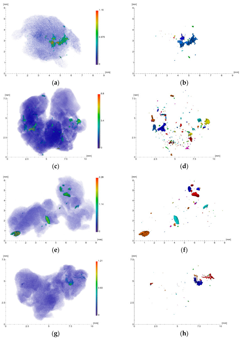
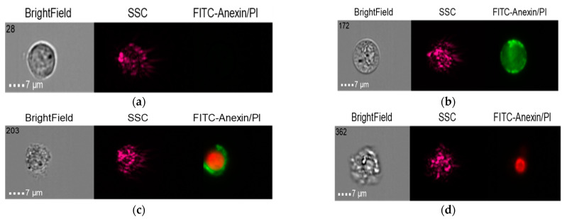
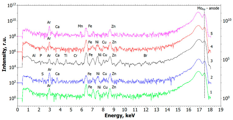
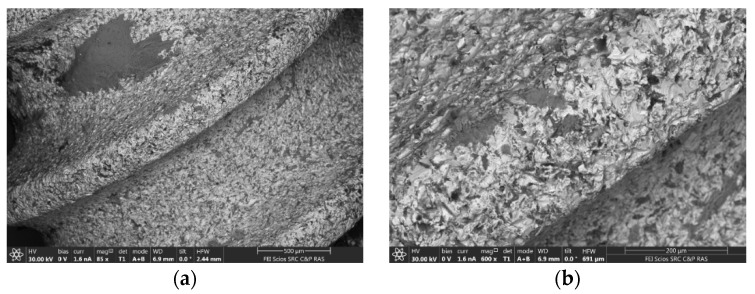
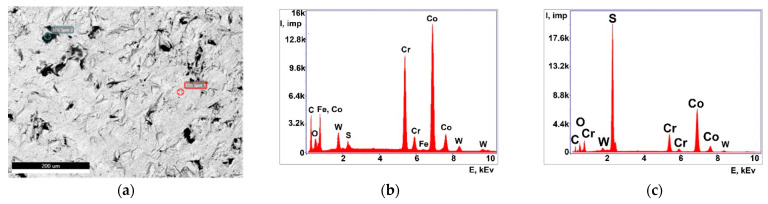
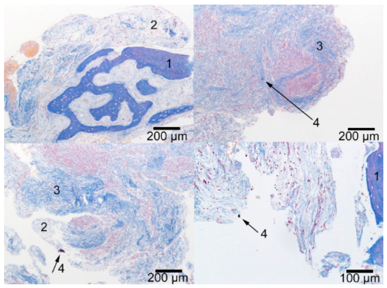
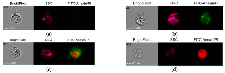
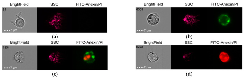
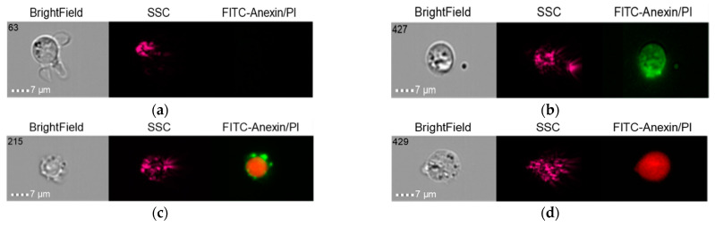
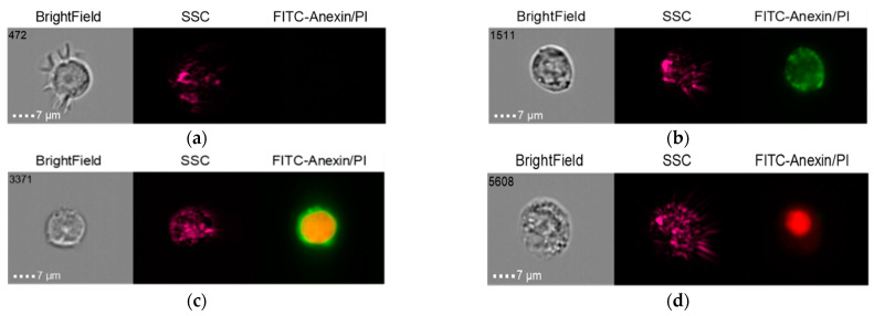
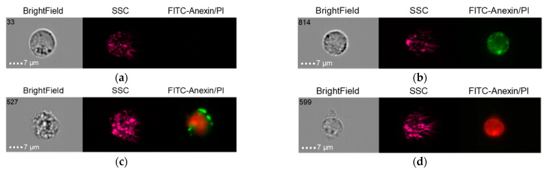
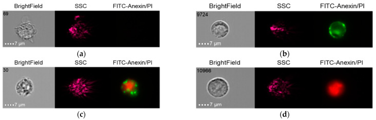
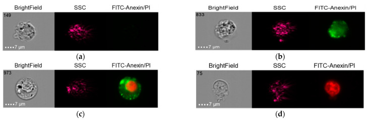
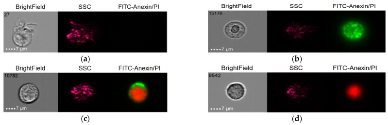
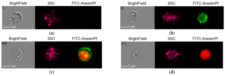
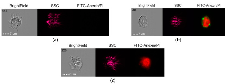
The role of metallic nano- and microparticles in the development of inflammation has not yet been investigated. Soft tissue biopsy specimens of the bone bed taken during surgical revisions, as well as supernatants obtained from the surface of the orthopedic structures and dental implants (control), were examined. Investigations were performed using X-ray microtomography, X-ray fluorescence analysis, and scanning electron microscopy. Histological studies of the bone bed tissues were performed. Nanoscale and microscale metallic particles were identified as participants in the inflammatory process in tissues. Supernatants containing nanoscale particles were obtained from the surfaces of 20 units of new dental implants. Early and late apoptosis and necrosis of immunocompetent cells after co-culture and induction by lipopolysaccharide and human venous blood serum were studied in an experiment with staging on the THP-1 (human monocytic) cell line using visualizing cytometry. As a result, it was found that nano- and microparticles emitted from the surface of the oxide layer of medical devices impregnated soft tissue biopsy specimens. By using different methods to analyze the cell-molecule interactions of nano- and microparticles both from a clinical perspective and an experimental research perspective, the possibility of forming a chronic immunopathological endogenous inflammatory process with an autoimmune component in the tissues was revealed.
金属纳米和微米颗粒在炎症发展中的作用尚未得到研究。在手术翻修过程中从骨床采集的软组织活检标本,以及从骨科结构和牙科植入物(对照)表面获得的上清液,都进行了检查。使用 X 射线微断层扫描、X 射线荧光分析和扫描电子显微镜进行了研究。对骨床组织进行了组织学研究。纳米级和微米级金属颗粒被鉴定为组织中炎症过程的参与者。从 20 个新的牙科植入物表面获得了含有纳米级颗粒的上清液。通过在 THP-1(人单核细胞)细胞系上进行分期实验,使用可视化细胞术研究了共培养和脂多糖及人静脉血清诱导后免疫活性细胞的早期和晚期细胞凋亡和坏死。结果发现,从涂有氧化物层的医疗器械表面发射出纳米和微米颗粒,渗透到软组织活检标本中。通过使用不同的方法从临床和实验研究的角度分析纳米和微米颗粒的细胞-分子相互作用,揭示了在组织中形成具有自身免疫成分的慢性免疫病理内源性炎症过程的可能性。