Max Planck Institute of Biochemistry, Martinsried, Germany.
Fondazione Human Technopole, Milano, Italy.
Nat Commun. 2021 Sep 10;12(1):5364. doi: 10.1038/s41467-021-25413-w.
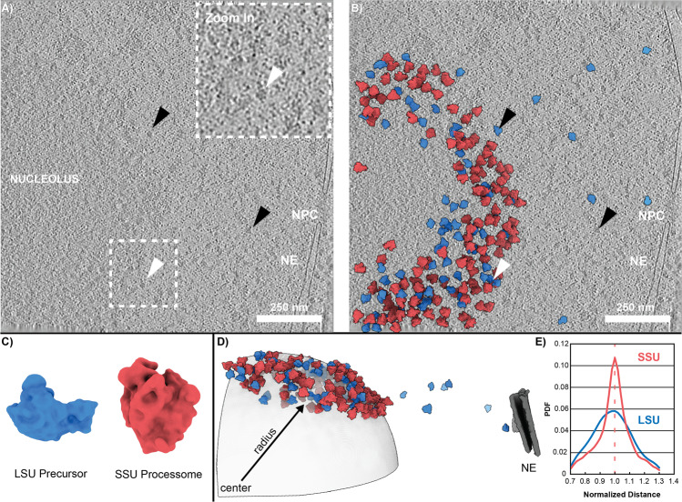
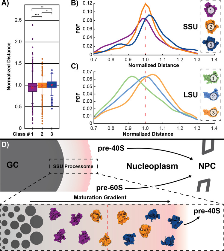
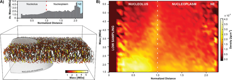
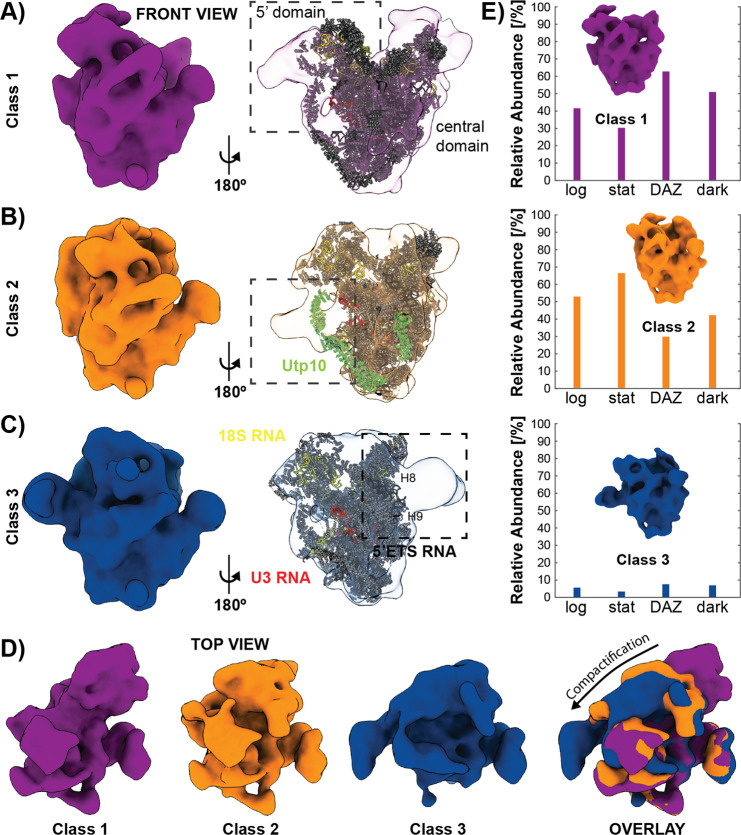
Ribosomes comprise a large (LSU) and a small subunit (SSU) which are synthesized independently in the nucleolus before being exported into the cytoplasm, where they assemble into functional ribosomes. Individual maturation steps have been analyzed in detail using biochemical methods, light microscopy and conventional electron microscopy (EM). In recent years, single particle analysis (SPA) has yielded molecular resolution structures of several pre-ribosomal intermediates. It falls short, however, of revealing the spatiotemporal sequence of ribosome biogenesis in the cellular context. Here, we present our study on native nucleoli in Chlamydomonas reinhardtii, in which we follow the formation of LSU and SSU precursors by in situ cryo-electron tomography (cryo-ET) and subtomogram averaging (STA). By combining both positional and molecular data, we reveal gradients of ribosome maturation within the granular component (GC), offering a new perspective on how the liquid-liquid-phase separation of the nucleolus supports ribosome biogenesis.
核糖体由一个大亚基(LSU)和一个小亚基(SSU)组成,它们在核仁中独立合成,然后被输出到细胞质中,在那里它们组装成功能核糖体。使用生化方法、光显微镜和传统电子显微镜(EM)已经详细分析了各个成熟步骤。近年来,单颗粒分析(SPA)已经获得了几个前核糖体中间产物的分子分辨率结构。然而,它无法揭示细胞环境中核糖体生物发生的时空顺序。在这里,我们展示了我们在莱茵衣藻中的天然核仁的研究,我们通过原位冷冻电子断层扫描(cryo-ET)和亚断层平均(STA)来跟踪 LSU 和 SSU 前体的形成。通过结合位置和分子数据,我们揭示了颗粒成分(GC)中核糖体成熟的梯度,为核仁的液-液相分离如何支持核糖体生物发生提供了新的视角。