Division of Cardiovascular Medicine, 300 Pasteur Drive, Falk CVRC, Stanford, CA, 94305, USA.
Research Center for Intelligent Computing Platforms, Zhejiang Laboratory, Hangzhou, 311121, China.
Nat Commun. 2023 Feb 15;14(1):847. doi: 10.1038/s41467-023-36518-9.
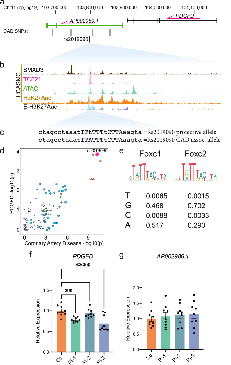
Genome wide association studies for coronary artery disease (CAD) have identified a risk locus at 11q22.3. Here, we verify with mechanistic studies that rs2019090 and PDGFD represent the functional variant and gene at this locus. Further, FOXC1/C2 transcription factor binding at rs2019090 is shown to promote PDGFD transcription through the CAD promoting allele. With single cell transcriptomic and histology studies with Pdgfd knockdown in an SMC lineage tracing male atherosclerosis mouse model we find that Pdgfd promotes expansion, migration, and transition of SMC lineage cells to the chondromyocyte phenotype. Pdgfd also increases adventitial fibroblast and pericyte expression of chemokines and leukocyte adhesion molecules, which is linked to plaque macrophage recruitment. Despite these changes there is no effect of Pdgfd deletion on overall plaque burden. These findings suggest that PDGFD mediates CAD risk by promoting deleterious phenotypic changes in SMC, along with an inflammatory response that is primarily focused in the adventitia.
全基因组关联研究发现,冠状动脉疾病(CAD)的风险位于 11q22.3。在这里,我们通过机制研究证实 rs2019090 和 PDGFD 是该基因座的功能性变体和基因。此外,FOXC1/C2 转录因子在 rs2019090 处的结合被证明可以通过 CAD 促进等位基因促进 PDGFD 转录。通过单细胞转录组学和组织学研究,在 SMC 谱系追踪的雄性动脉粥样硬化小鼠模型中敲低 Pdgfd,我们发现 Pdgfd 促进 SMC 谱系细胞的扩张、迁移和向软骨母细胞表型的转变。Pdgfd 还增加了外膜成纤维细胞和周细胞中趋化因子和白细胞黏附分子的表达,这与斑块内巨噬细胞的募集有关。尽管发生了这些变化,但 Pdgfd 缺失对斑块总负担没有影响。这些发现表明,PDGFD 通过促进 SMC 中有害的表型变化以及主要集中在外膜的炎症反应来介导 CAD 风险。