Department of Pathology, the Third Affiliated Hospital, Guangzhou Medical University, Guangzhou, China.
Guangdong Provincial Key Laboratory of Major Obstetric Diseases, Guangzhou, China.
Diagn Pathol. 2023 Feb 16;18(1):23. doi: 10.1186/s13000-023-01308-w.
The precise grading and characterization of cervical intraepithelial neoplasia (CIN) has been the focus of pathologists for a long time. This study aimed to explore known strategies for the grading of CINs.
After routine H&E review, 85 lesions graded CIN 1, 2, or 3 were investigated primarily by HPV RNAscope to detect HR-HPV and LR-HPV, in combination with an HPV-DNA test and P16/Ki67 immunohistochemistry (IHC). Then, the 85 cases were divided into a control group (49 cases) and a test group (36 cases). The former consisted of cases with consistency between morphology, HPV DNA detection and P16/Ki67 IHC. We used them to evaluate HPV RNA distribution patterns in CINs of different grades. The latter were ambiguous cases in which pathologists could not confirm the diagnosis because of inconsistencies between morphology, HPV DNA detection and P16/Ki67 IHC. We reassessed them by comparison to the pattern in the control group.
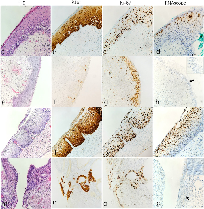
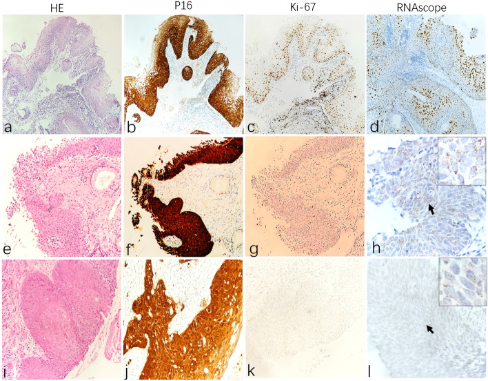
The expression patterns of HPV mRNA signals were different in different CIN lesions. LSIL/CIN1 lesions were mostly expressed in superficial epithelium with diffuse clustered nuclear or cytoplasmic staining; HSIL/CIN2 were characterised by nuclear/cytoplasmic punctate or diffuse cluster nuclear staining in the mid-surface layer, and scattered nuclear/cytoplasmic punctate staining in basal and parabasal cells; whereas HSIL/CIN3 showed full-thickness nucleus/cytoplasmic scattered staining with a punctate pattern. According to the staining pattern, we corrected the diagnosis of 22 cases (22/36, 61.1%).
Because of its distinct location pattern, HPV RNAscope has obvious advantages over the HPV-DNA test, and combined with P16/Ki67 IHC, it can help pathologists correctly grade CIN. In addition, it can effectively discriminate true CIN from normal or CIN mimic lesions, such as immature squamous metaplasia, atrophy, and inflammatory/reactive changes. Therefore, HPV RNAscope is a valuable auxiliary diagnostic test to avoid the overtreatment and undertreatment of CIN lesions.
长期以来,宫颈上皮内瘤变(CIN)的准确分级和特征描述一直是病理学家关注的焦点。本研究旨在探讨 CIN 分级的已知策略。
在常规 H&E 复查后,主要通过 HPV RNAscope 检测 HR-HPV 和 LR-HPV,结合 HPV-DNA 检测和 P16/Ki67 免疫组化(IHC)对 85 例 CIN1、2 或 3 级病变进行研究。然后,将 85 例病例分为对照组(49 例)和实验组(36 例)。前者由形态学、HPV DNA 检测和 P16/Ki67 IHC 一致的病例组成。我们用它们来评估不同分级 CIN 中 HPV RNA 分布模式。后者为形态学、HPV DNA 检测和 P16/Ki67 IHC 不一致,病理医生无法确诊的模棱两可病例。我们通过与对照组进行比较重新评估这些病例。
HPV mRNA 信号的表达模式在不同的 CIN 病变中不同。LSIL/CIN1 病变主要在上皮层表达,呈弥漫性簇状核或细胞质染色;HSIL/CIN2 病变的特点是中表层核/细胞质点状或弥漫簇状核染色,基底和副基底细胞散在点状核/细胞质染色;而 HSIL/CIN3 表现为全层核/细胞质点状散在染色。根据染色模式,我们纠正了 22 例(22/36,61.1%)的诊断。
由于其位置模式明显,HPV RNAscope 明显优于 HPV-DNA 检测,结合 P16/Ki67 IHC,有助于病理医生正确分级 CIN。此外,它可以有效地将真正的 CIN 与正常或 CIN 模拟病变(如不成熟的鳞状化生、萎缩和炎症/反应性改变)区分开来。因此,HPV RNAscope 是一种有价值的辅助诊断试验,可以避免 CIN 病变的过度治疗和治疗不足。