Department of Emergency, First Affiliated Hospital of Wenzhou Medical University, Shangcai Road, Ouhai District, Wenzhou, 325000, Zhejiang, China.
Department of Critical Care Medicine, Xijing Hospital, Xi'an, 710000, Shaanxi, China.
Mol Med. 2023 Feb 21;29(1):25. doi: 10.1186/s10020-023-00618-5.
Dendritic cell (DC) dysfunction plays a central role in sepsis-induced immunosuppression. Recent research has indicated that collective mitochondrial fragmentation contributes to the dysfunction of immune cells observed during sepsis. PTEN-induced putative kinase 1 (PINK1) has been characterized as a guide for impaired mitochondria that can keep mitochondrial homeostasis. However, its role in the function of DCs during sepsis and the related mechanisms remain obscure. In our study, we elucidated the effect of PINK1 on DC function during sepsis and its underlying mechanism of action.
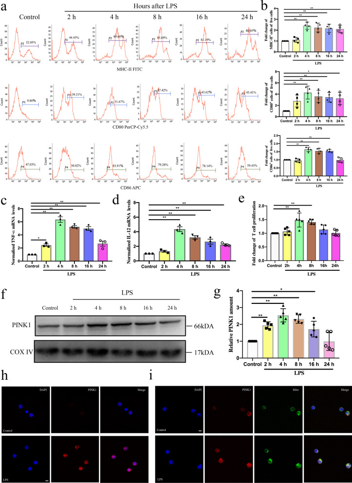
Cecal ligation and puncture (CLP) surgery and lipopolysaccharide (LPS) treatment were used as in vivo and in vitro sepsis models, respectively.
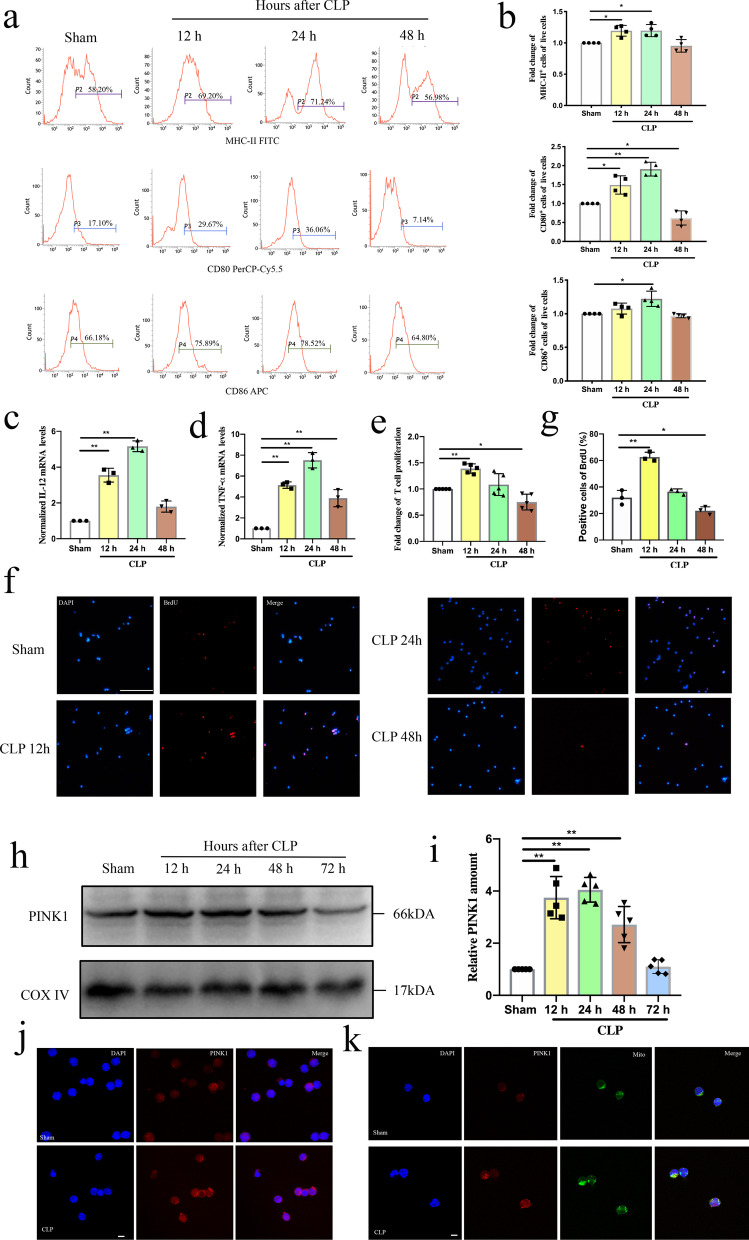
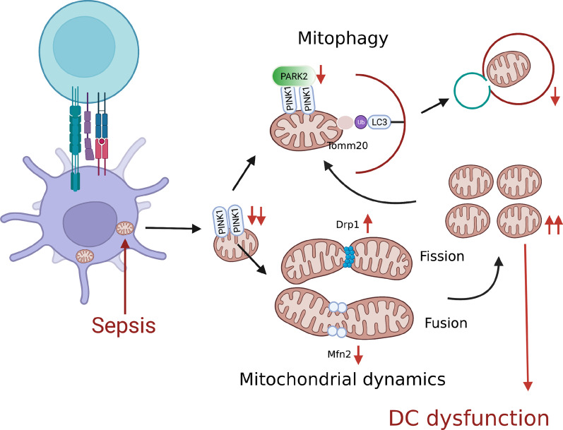
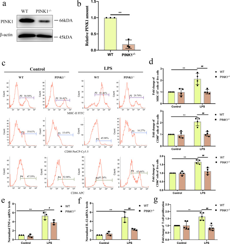
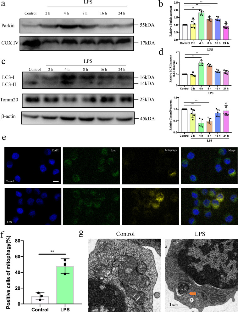
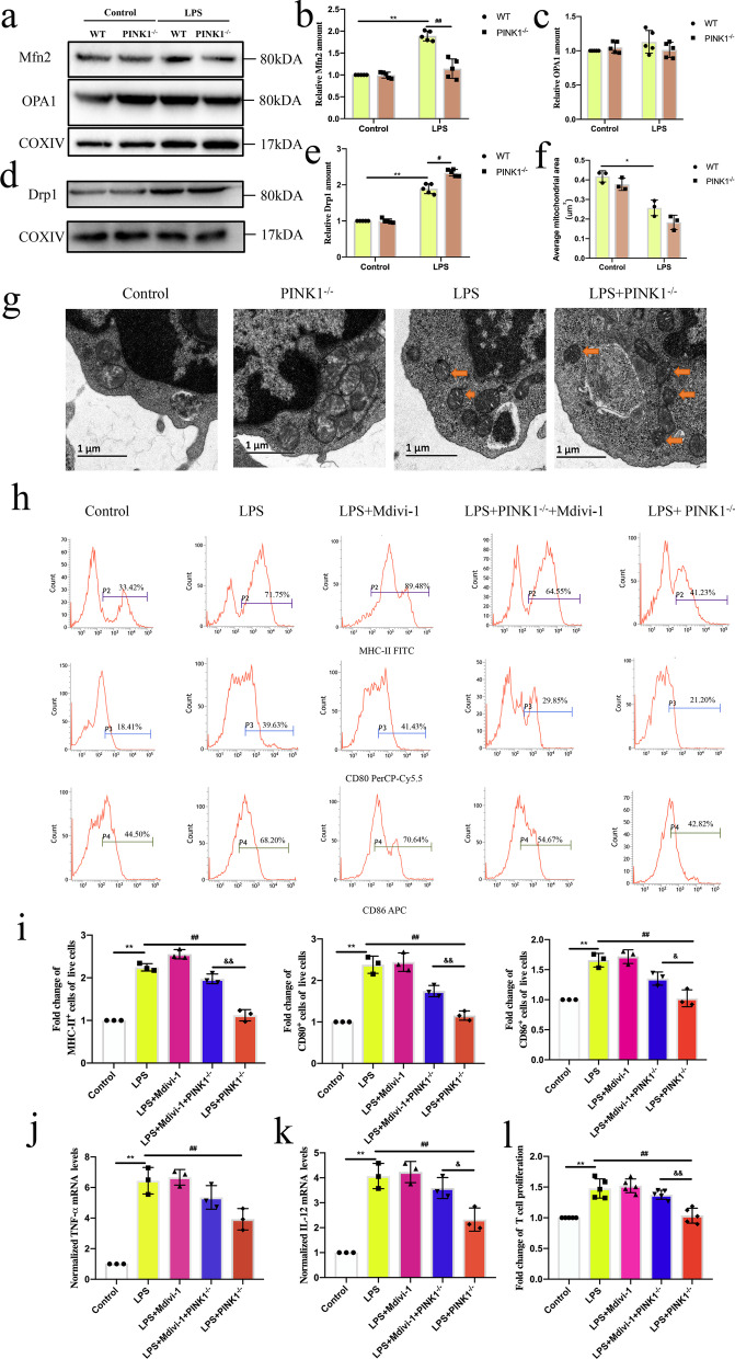
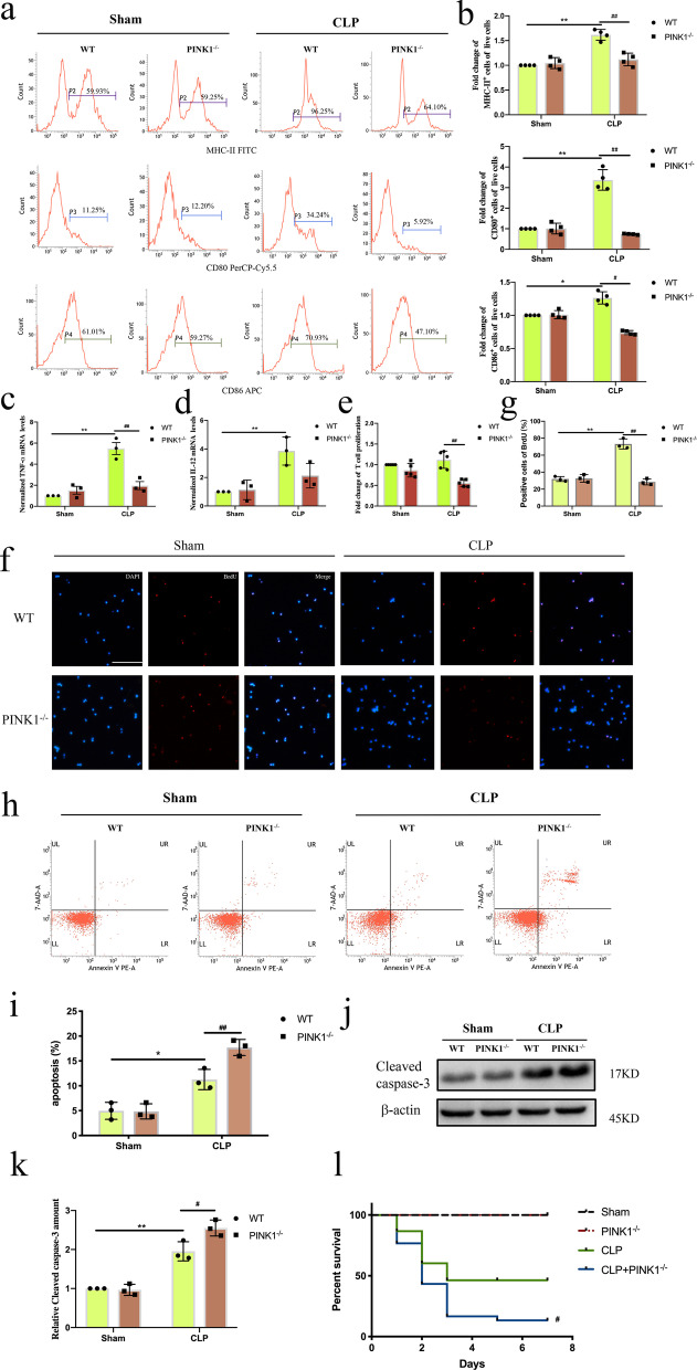
We found that changes in mitochondrial PINK1 expression of DCs paralleled changes in DC function during sepsis. The ratio of DCs expressing MHC-II, CD86, and CD80, the mRNAs level of dendritic cells expressing TNF-α and IL-12, and the level of DC-mediated T-cell proliferation were all decreased, both in vivo and in vitro during sepsis, when PINK1 was knocked out. This suggested that PINK1 knockout prevented the function of DCs during sepsis. Furthermore, PINK1 knockout inhibited Parkin RBR E3 ubiquitin protein (Parkin)-dependent mitophagy and enhanced dynamin-related protein 1 (Drp1)-related mitochondrial fission, and the negative effects of PINK1 knockout on DC function following LPS treatment were reversed by Parkin activation and Drp1 inhibitor. Knockout of PINK1 also increased apoptosis of DCs and the mortality of CLP mice.
Our results indicated that PINK1 protected against DC dysfunction during sepsis through the regulation of mitochondrial quality control.
树突状细胞(DC)功能障碍在脓毒症引起的免疫抑制中起核心作用。最近的研究表明,集体线粒体碎片化导致脓毒症期间观察到的免疫细胞功能障碍。PTEN 诱导的假定激酶 1(PINK1)已被描述为指导受损线粒体的指南,可维持线粒体的动态平衡。然而,其在脓毒症期间 DC 功能中的作用及其相关机制尚不清楚。在本研究中,我们阐明了 PINK1 在脓毒症期间对 DC 功能的影响及其作用机制。
使用盲肠结扎和穿孔(CLP)手术和脂多糖(LPS)处理分别作为体内和体外脓毒症模型。
我们发现,DC 中线粒体 PINK1 表达的变化与脓毒症期间 DC 功能的变化平行。在体内和体外脓毒症期间,当敲除 PINK1 时,表达 MHC-II、CD86 和 CD80 的 DC 比例、表达 TNF-α 和 IL-12 的 DCs 的 mRNA 水平以及 DC 介导的 T 细胞增殖水平均降低。这表明 PINK1 敲除可防止脓毒症期间 DC 的功能。此外,PINK1 敲除抑制 Parkin RBR E3 泛素蛋白(Parkin)依赖性线粒体自噬,并增强与 dynamin 相关蛋白 1(Drp1)相关的线粒体裂变,而 LPS 处理后 PINK1 敲除对 DC 功能的负效应可通过 Parkin 激活和 Drp1 抑制剂逆转。敲除 PINK1 还增加了 DC 的凋亡和 CLP 小鼠的死亡率。
我们的结果表明,PINK1 通过调节线粒体质量控制来防止脓毒症期间 DC 功能障碍。