• 文献检索
  • 文档翻译
  • 深度研究
  • 学术资讯
  • Suppr Zotero 插件Zotero 插件
  • 邀请有礼
  • 套餐&价格
  • 历史记录
应用&插件
Suppr Zotero 插件Zotero 插件浏览器插件Mac 客户端Windows 客户端微信小程序
定价
高级版会员购买积分包购买API积分包
服务
文献检索文档翻译深度研究API 文档MCP 服务
关于我们
关于 Suppr公司介绍联系我们用户协议隐私条款
关注我们

Suppr 超能文献

核心技术专利:CN118964589B侵权必究
粤ICP备2023148730 号-1Suppr @ 2026

文献检索

告别复杂PubMed语法,用中文像聊天一样搜索,搜遍4000万医学文献。AI智能推荐,让科研检索更轻松。

立即免费搜索

文件翻译

保留排版,准确专业,支持PDF/Word/PPT等文件格式,支持 12+语言互译。

免费翻译文档

深度研究

AI帮你快速写综述,25分钟生成高质量综述,智能提取关键信息,辅助科研写作。

立即免费体验

间质真皮淋巴细胞浸润内CD34表达缺失是硬斑病诊断的一个有用线索。

Loss of CD34 Expression within an Interstitial Dermal Lymphoid Cell Infiltrate Is a Helpful Clue to the Diagnosis of Morphea.

作者信息

Daruish Maged, Zidan Anoud, Greenblatt Danielle T, Stefanato Catherine M

机构信息

Department of Dermatopathology, St John's Institute of Dermatology, Guy's and St Thomas' NHS Foundation Trust, London SE1 7EH, UK.

Pediatric Dermatology, St John's Institute of Dermatology, Guy's and St Thomas' NHS Foundation Trust, London SE1 9RT, UK.

出版信息

Dermatopathology (Basel). 2023 Feb 2;10(1):70-74. doi: 10.3390/dermatopathology10010010.

DOI:10.3390/dermatopathology10010010
PMID:36810568
原文链接:https://pmc.ncbi.nlm.nih.gov/articles/PMC9944576/
Abstract

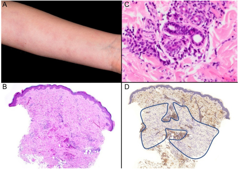
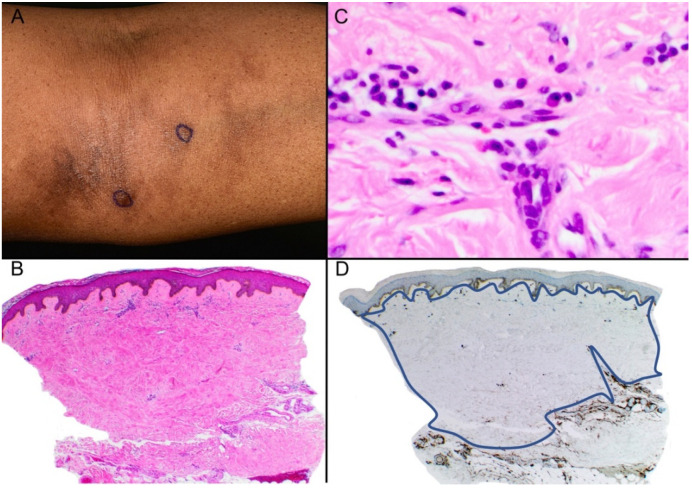
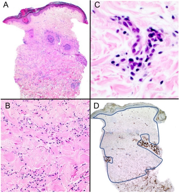
A dermal interstitial lymphocytic infiltrate may represent a diagnostic challenge, particularly if the clinical history is not provided. We present three cases within the histological spectrum of morphea in which the immunohistochemical marker CD34 was helpful in confirming the diagnosis.

摘要

皮肤间质淋巴细胞浸润可能带来诊断挑战,尤其是在未提供临床病史的情况下。我们展示了三例硬斑病组织学范围内的病例,其中免疫组化标志物CD34有助于确诊。

https://cdn.ncbi.nlm.nih.gov/pmc/blobs/dc9a/9944576/d3c43586fca0/dermatopathology-10-00010-g003.jpg
https://cdn.ncbi.nlm.nih.gov/pmc/blobs/dc9a/9944576/6dfef9275610/dermatopathology-10-00010-g001.jpg
https://cdn.ncbi.nlm.nih.gov/pmc/blobs/dc9a/9944576/74eca95443ec/dermatopathology-10-00010-g002.jpg
https://cdn.ncbi.nlm.nih.gov/pmc/blobs/dc9a/9944576/d3c43586fca0/dermatopathology-10-00010-g003.jpg
https://cdn.ncbi.nlm.nih.gov/pmc/blobs/dc9a/9944576/6dfef9275610/dermatopathology-10-00010-g001.jpg
https://cdn.ncbi.nlm.nih.gov/pmc/blobs/dc9a/9944576/74eca95443ec/dermatopathology-10-00010-g002.jpg
https://cdn.ncbi.nlm.nih.gov/pmc/blobs/dc9a/9944576/d3c43586fca0/dermatopathology-10-00010-g003.jpg

相似文献

1
Loss of CD34 Expression within an Interstitial Dermal Lymphoid Cell Infiltrate Is a Helpful Clue to the Diagnosis of Morphea.间质真皮淋巴细胞浸润内CD34表达缺失是硬斑病诊断的一个有用线索。
Dermatopathology (Basel). 2023 Feb 2;10(1):70-74. doi: 10.3390/dermatopathology10010010.
2
Interstitial mycosis fungoides, a variant of mycosis fungoides resembling granuloma annulare and inflammatory morphea.间质性蕈样肉芽肿,是蕈样肉芽肿的一种变体,类似环状肉芽肿和炎症性硬斑病。
J Cutan Pathol. 2002 Mar;29(3):135-41. doi: 10.1034/j.1600-0560.2002.290302.x.
3
CD34 Staining as a Useful Tool in Disorders of Collagen Degeneration.CD34染色作为胶原变性疾病的一种有用工具。
Am J Dermatopathol. 2022 Feb 1;44(2):103-105. doi: 10.1097/DAD.0000000000002002.
4
Morphea limited to the superficial reticular dermis: an underrecognized histologic phenomenon.局限性硬斑病仅限于浅表网状真皮层:一种未被充分认识的组织学现象。
Am J Dermatopathol. 1999 Aug;21(4):315-9. doi: 10.1097/00000372-199908000-00001.
5
Elastic fiber pattern in scleroderma/morphea.硬皮病/局限性硬皮病中的弹性纤维模式
J Cutan Pathol. 2009 Sep;36(9):952-7. doi: 10.1111/j.1600-0560.2009.01201.x.
6
CD34 stromal expression is inversely proportional to smooth muscle actin expression and extent of morphea.CD34在基质中的表达与平滑肌肌动蛋白的表达及硬斑病的范围呈负相关。
J Eur Acad Dermatol Venereol. 2018 Dec;32(12):2208-2216. doi: 10.1111/jdv.15120. Epub 2018 Jul 6.
7
Histopathologic spectrum of morphea: a single-center retrospective study.硬斑病的组织病理学谱:一项单中心回顾性研究。
Dermatol Reports. 2024 Feb 6;16(3):9915. doi: 10.4081/dr.2024.9915. eCollection 2024 Sep 2.
8
Decreased expression of the human progenitor cell antigen (CD34) in morphea.局限性硬皮病中人类祖细胞抗原(CD34)表达降低。
Am J Dermatopathol. 1995 Oct;17(5):471-5. doi: 10.1097/00000372-199510000-00007.
9
Immunohistochemical study of epidermal Langerhans cells and dermal dendritic cells in benign and malignant skin lesions characterized by a dermal lymphoid infiltrate consisting either of B-cells or T-cells.对以B细胞或T细胞组成的真皮淋巴细胞浸润为特征的良性和恶性皮肤病变中的表皮朗格汉斯细胞和真皮树突状细胞进行免疫组织化学研究。
Virchows Arch A Pathol Anat Histopathol. 1987;411(4):337-43. doi: 10.1007/BF00713378.
10
A Case of Circumscribed Scalp Morphea with Perineural Lymphocytes on Pathology.一例病理检查显示有神经周淋巴细胞的局限性头皮硬斑病病例。
Skin Appendage Disord. 2017 Oct;3(4):175-178. doi: 10.1159/000471855. Epub 2017 Apr 29.

引用本文的文献

1
Paradoxical morphea-like reaction after initiation of dupilumab for nodular prurigo.度普利尤单抗治疗结节性痒疹开始后出现的矛盾性硬斑病样反应。
An Bras Dermatol. 2025 Aug 14;100(5):501193. doi: 10.1016/j.abd.2025.501193.
2
[Clues in dermatopathological diagnostics].[皮肤病理学诊断中的线索]
Pathologie (Heidelb). 2025 Mar;46(2):81-89. doi: 10.1007/s00292-024-01400-y. Epub 2024 Dec 9.

本文引用的文献

1
Interstitial Mycosis Fungoides: An Unusual Mimic of Interstitial Granuloma Annulare Not to Miss.间质性蕈样霉菌病:一种不可漏诊的不寻常的环状肉芽肿样表现。
Case Rep Dermatol Med. 2022 Sep 5;2022:3506738. doi: 10.1155/2022/3506738. eCollection 2022.
2
CD34 Staining as a Useful Tool in Disorders of Collagen Degeneration.CD34染色作为胶原变性疾病的一种有用工具。
Am J Dermatopathol. 2022 Feb 1;44(2):103-105. doi: 10.1097/DAD.0000000000002002.
3
Histopathologic Spectrum of Morphea.硬斑病的组织病理学谱。
Am J Dermatopathol. 2021 Jan 1;43(1):1-8. doi: 10.1097/DAD.0000000000001662.
4
Histopathology of morphea: Sensitivity of various named signs, a retrospective study.硬斑病的组织病理学:各种命名性征象的敏感性,一项回顾性研究。
Indian J Pathol Microbiol. 2020 Oct-Dec;63(4):600-603. doi: 10.4103/IJPM.IJPM_67_20.
5
CD34 Staining in Disorders of Collagen Degeneration Other Than Morphea.局限性硬皮病以外的胶原变性疾病中的CD34染色
Am J Dermatopathol. 2020 Aug;42(8):623-624. doi: 10.1097/DAD.0000000000001584.
6
CD34 stromal expression is inversely proportional to smooth muscle actin expression and extent of morphea.CD34在基质中的表达与平滑肌肌动蛋白的表达及硬斑病的范围呈负相关。
J Eur Acad Dermatol Venereol. 2018 Dec;32(12):2208-2216. doi: 10.1111/jdv.15120. Epub 2018 Jul 6.
7
Decreased expression of the human progenitor cell antigen (CD34) in morphea.局限性硬皮病中人类祖细胞抗原(CD34)表达降低。
Am J Dermatopathol. 1995 Oct;17(5):471-5. doi: 10.1097/00000372-199510000-00007.
8
The histologic spectrum of mycosis fungoides/Sézary syndrome (cutaneous T-cell lymphoma). A review of 222 biopsies, including newly described patterns and the earliest pathologic changes.蕈样肉芽肿/赛塞里综合征(皮肤T细胞淋巴瘤)的组织学谱。对222例活检的回顾,包括新描述的模式和最早的病理变化。
Am J Surg Pathol. 1994 Jul;18(7):645-67. doi: 10.1097/00000478-199407000-00001.
9
CD34+ spindle-shaped cells selectively disappear from the skin lesion of scleroderma.CD34+梭形细胞从硬皮病的皮肤病变中选择性消失。
Arch Dermatol. 1994 May;130(5):593-7.