Huang Cicheng, Zhan Chenao, Hu Yiqi, Yin Ting, Grimm Robert, Ai Tao
Center of Stomatology, Tongji Hospital, Tongji Medical College, Huazhong University of Science and Technology, Wuhan, China.
Department of Radiology, Tongji Hospital, Tongji Medical College, Huazhong University of Science and Technology, Wuhan, China.
Quant Imaging Med Surg. 2023 Feb 1;13(2):735-746. doi: 10.21037/qims-22-475. Epub 2023 Jan 2.
Histogram analysis of the diffusion-weighted imaging (DWI) parameters is widely used to differentiate the breast lesions. However, histogram analysis of the diffusion-kurtosis imaging (DKI) parameters for the single-shot echo-planar imaging (ss-EPI) and readout-segmented echo planar imaging (rs-EPI) sequences has not been compared in breast cancer. Thus, this study is to investigate the diagnostic accuracy and reliability of the histogram parameters derived from the rs-EPI and ss-EPI sequences of DKI parameters in distinguishing between the benign and malignant breast lesions.
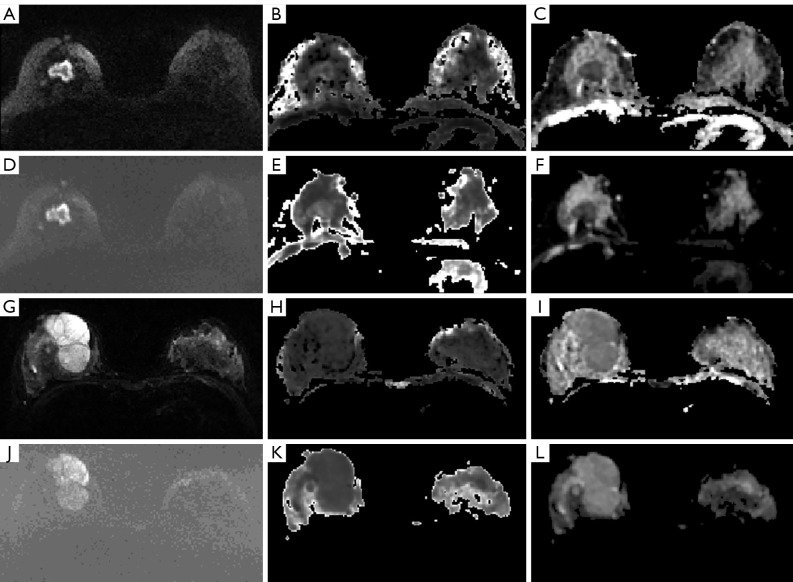
This single-center, retrospective cohort study enrolled 205 consecutive patients with breast lesions (65 benign and 140 malignant). The patients underwent breast magnetic resonance imaging (MRI) with a 3T scanner using the rs-EPI and ss-EPI sequences with 4 b values (0, 50, 1,000, and 2,000 s/mm). The regions of interest (ROIs) were manually delineated for all the lesion images from both the sequences, and the histogram parameters were extracted from the apparent diffusion coefficient (ADC) and apparent diffusional kurtosis (K) maps. Statistical analysis was performed using the Kolmogorov-Smirnov test, the student's test, and the receiver operating characteristic (ROC) curves.
The mean, 25th, 50th, 75th, and 100th percentiles, skewness, and kurtosis values derived from apparent diffusion for non-Gaussian distribution (D) and K maps showed good or excellent intra-observer agreement (ICC: 0.695 to 0.863).The mean and the 25th, 50th, 75th, and 100th percentile values for D were significantly lower and the mean and the 25th, 50th, 75th, and 100th percentile values for K were significantly higher in the malignant breast lesions compared with those in the benign breast lesions for both the rs-EPI and ss-EPI sequences (all P<0.05). The majority of the histogram K and D parameters (except skewness and kurtosis) for the benign and malignant lesions showed significant differences between the ss-EPI and the rs-EPI sequences (P<0.05). ROC curve analysis showed that the AUC values for the 75th percentile of K (0.854 for rs-EPI, 0.844 for ss-EPI) and the 25th percentile of D (0.866 for rs-EPI, 0.858 for ss-EPI) were highest for both DKI sequences. The diagnostic performance of the rs-EPI sequence was better than the ss-EPI sequence for all the histogram parameters except the skewness value of D.
Histogram parameters from the rs-EPI sequence were more reliable and accurate in differentiating malignant and benign breast lesions than those from the ss-EPI sequence.
扩散加权成像(DWI)参数的直方图分析广泛用于鉴别乳腺病变。然而,尚未对乳腺癌单次激发回波平面成像(ss-EPI)序列和读出分段回波平面成像(rs-EPI)序列的扩散峰度成像(DKI)参数进行直方图分析比较。因此,本研究旨在探讨DKI参数的rs-EPI和ss-EPI序列得出的直方图参数在鉴别乳腺良恶性病变中的诊断准确性和可靠性。
本单中心回顾性队列研究纳入了205例连续的乳腺病变患者(65例良性病变和140例恶性病变)。患者使用3T扫描仪,采用rs-EPI和ss-EPI序列,4个b值(0、50、1000和2000 s/mm²)进行乳腺磁共振成像(MRI)检查。对两个序列的所有病变图像手动勾画感兴趣区(ROI),并从表观扩散系数(ADC)和表观扩散峰度(K)图中提取直方图参数。采用Kolmogorov-Smirnov检验、t检验和受试者工作特征(ROC)曲线进行统计分析。
非高斯分布的表观扩散(D)图和K图得出的均值、第25、50、75和100百分位数、偏度和峰度值显示观察者内一致性良好或优秀(ICC:0.695至0.863)。对于rs-EPI和ss-EPI序列,恶性乳腺病变的D均值及第25、50、75和100百分位数均显著低于良性乳腺病变,而K均值及第25、50、75和100百分位数均显著高于良性乳腺病变(所有P<0.05)。良性和恶性病变的大多数直方图K和D参数(偏度和峰度除外)在ss-EPI和rs-EPI序列之间存在显著差异(P<0.05)。ROC曲线分析显示,对于两个DKI序列,K的第75百分位数(rs-EPI为0.854,ss-EPI为0.844)和D的第25百分位数(rs-EPI为0.866,ss-EPI为0.858)的AUC值最高。除D的偏度值外,rs-EPI序列在所有直方图参数方面的诊断性能均优于ss-EPI序列。
rs-EPI序列的直方图参数在鉴别乳腺良恶性病变方面比ss-EPI序列更可靠、准确。