Tian Meng, Zeng Guodong, Tappeiner Christoph, Zinkernagel Martin S, Wolf Sebastian, Munk Marion R
Beijing Tongren Eye Center, Beijing Tongren Hospital, Capital Medical University, Beijing, China.
Department of Ophthalmology, Inselspital, Bern University Hospital, University of Bern, Bern, Switzerland.
Front Med (Lausanne). 2022 May 2;9:853315. doi: 10.3389/fmed.2022.853315. eCollection 2022.
To compare indocyanine green angiography (ICGA) and swept-source wide-field optical coherence tomography angiography (SS-OCTA) for the assessment of patients with posterior uveitis.
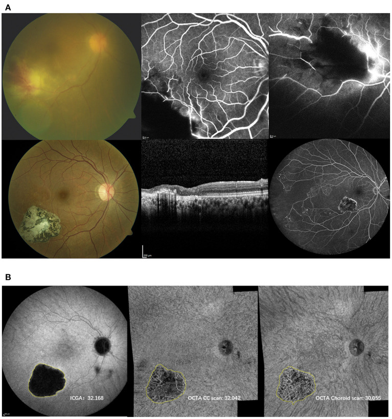
SS-OCTA montage images of 5 x 12 x 12 mm or 2 x 15 x 9 mm, covering ~70-90 degree of the retina of consecutive patients with posterior uveitis were acquired. The choriocapillaries and choroidal slabs were compared to findings on ICGA.
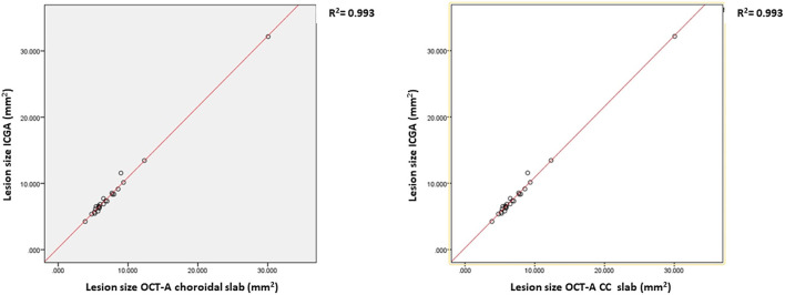
Sixty-eight eyes of 41 patients were included (mean age 47.2 ± 20.4 years; 58.5% female). In 23 (34%) lesions were visible on OCTA, but not discernable on ICGA. In turn, out of the 45 eyes with clearly discernable lesions on ICGA, 22 (49%) and 21 (47%) eyes showed no corresponding areas of flow deficit on OCTA in the CC and choroidal slab, respectively. Lesion size strongly correlated among ICGA and OCTA choriocapillaries- (CC) (r = 0.99, ≤ 0.0001) and choroidal slabs (r = 0.99, ≤ 0.0001), respectively. The mean lesion size on the late frames of ICGA (8.45 ± 5.47 mm) was larger compared to the lesion size on OCTA CC scan (7.98 ± 5.47 mm, ≤ 0.0001) and choroidal scan (7.69 ± 5.10 mm, = 0.002), respectively. The lesion size on OCTA CC scan was significantly larger than on the OCTA choroidal scan ( ≤ 0.0001).
SS-wide field OCTA may be a promising tool to assess posterior uveitis patients and may replace ICGA to a certain extent in the future.
比较吲哚菁绿血管造影(ICGA)和扫频源广角光学相干断层扫描血管造影(SS - OCTA)在评估后葡萄膜炎患者中的应用。
获取连续后葡萄膜炎患者视网膜约70 - 90度范围的5×12×12 mm或2×15×9 mm的SS - OCTA拼接图像。将脉络膜毛细血管和脉络膜层与ICGA检查结果进行比较。
纳入41例患者的68只眼(平均年龄47.2±20.4岁;女性占58.5%)。23只眼(34%)的病变在OCTA上可见,但在ICGA上无法分辨。反之,在ICGA上有清晰可辨病变的45只眼中,分别有22只眼(49%)和21只眼(47%)在OCTA的脉络膜毛细血管(CC)和脉络膜层中未显示相应的血流缺失区域。ICGA与OCTA的脉络膜毛细血管(CC)(r = 0.99,P≤0.0001)和脉络膜层(r = 0.99,P≤0.0001)的病变大小分别具有很强的相关性。ICGA晚期图像上的平均病变大小(8.45±5.47 mm)分别大于OCTA的CC扫描(7.98±5.47 mm,P≤0.0001)和脉络膜扫描(7.69±5.10 mm,P = 0.002)上的病变大小。OCTA的CC扫描上的病变大小显著大于OCTA的脉络膜扫描(P≤0.0001)。
SS - 广角OCTA可能是评估后葡萄膜炎患者的一种有前景的工具,未来可能在一定程度上取代ICGA。