Xia Yi, Zhang Xu, An Peng, Luo Junjie, Luo Yongting
Department of Nutrition and Health, China Agricultural University, Beijing 100193, China.
Int J Mol Sci. 2023 Feb 9;24(4):3483. doi: 10.3390/ijms24043483.
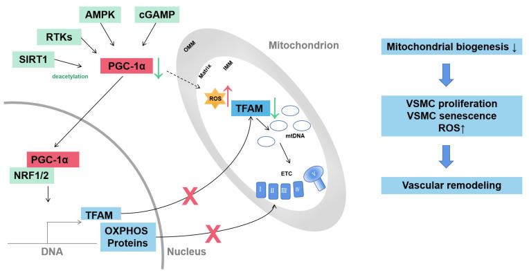
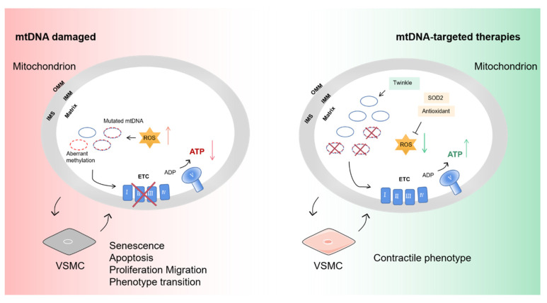
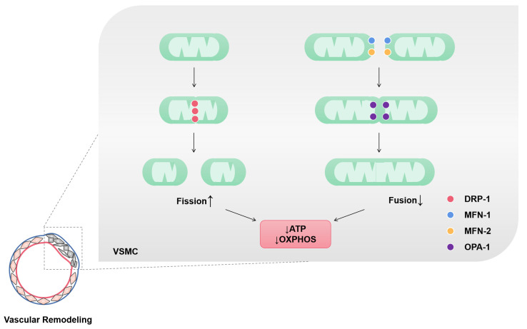
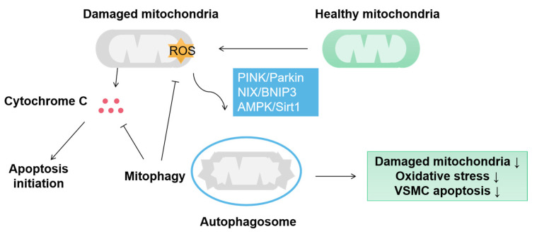
Vascular remodeling is a common pathological hallmark of many cardiovascular diseases. Vascular smooth muscle cells (VSMCs) are the predominant cell type lining the tunica media and play a crucial role in maintaining aortic morphology, integrity, contraction and elasticity. Their abnormal proliferation, migration, apoptosis and other activities are tightly associated with a spectrum of structural and functional alterations in blood vessels. Emerging evidence suggests that mitochondria, the energy center of VSMCs, participate in vascular remodeling through multiple mechanisms. For example, peroxisome proliferator-activated receptor-γ coactivator-1α (PGC-1α)-mediated mitochondrial biogenesis prevents VSMCs from proliferation and senescence. The imbalance between mitochondrial fusion and fission controls the abnormal proliferation, migration and phenotypic transformation of VSMCs. Guanosine triphosphate-hydrolyzing enzymes, including mitofusin 1 (MFN1), mitofusin 2 (MFN2), optic atrophy protein 1 (OPA1) and dynamin-related protein 1 (DRP1), are crucial for mitochondrial fusion and fission. In addition, abnormal mitophagy accelerates the senescence and apoptosis of VSMCs. PINK/Parkin and NIX/BINP3 pathways alleviate vascular remodeling by awakening mitophagy in VSMCs. Mitochondrial DNA (mtDNA) damage destroys the respiratory chain of VSMCs, resulting in excessive ROS production and decreased ATP levels, which are related to the proliferation, migration and apoptosis of VSMCs. Thus, maintaining mitochondrial homeostasis in VSMCs is a possible way to relieve pathologic vascular remodeling. This review aims to provide an overview of the role of mitochondria homeostasis in VSMCs during vascular remodeling and potential mitochondria-targeted therapies.
血管重塑是许多心血管疾病常见的病理特征。血管平滑肌细胞(VSMCs)是中膜的主要细胞类型,在维持主动脉形态、完整性、收缩和弹性方面起着至关重要的作用。它们的异常增殖、迁移、凋亡及其他活动与血管的一系列结构和功能改变密切相关。新出现的证据表明,VSMCs的能量中心线粒体通过多种机制参与血管重塑。例如,过氧化物酶体增殖物激活受体γ辅激活因子1α(PGC-1α)介导的线粒体生物合成可防止VSMCs增殖和衰老。线粒体融合与裂变之间的失衡控制着VSMCs的异常增殖、迁移和表型转化。包括线粒体融合蛋白1(MFN1)、线粒体融合蛋白2(MFN2)、视神经萎缩蛋白1(OPA1)和动力相关蛋白1(DRP1)在内的鸟苷三磷酸水解酶对线粒体融合和裂变至关重要。此外,异常的线粒体自噬加速了VSMCs的衰老和凋亡。PINK/Parkin和NIX/BINP3途径通过激活VSMCs中的线粒体自噬来减轻血管重塑。线粒体DNA(mtDNA)损伤破坏了VSMCs的呼吸链,导致活性氧过量产生和ATP水平降低,这与VSMCs的增殖、迁移和凋亡有关。因此,维持VSMCs中的线粒体稳态是缓解病理性血管重塑的一种可能途径。本综述旨在概述血管重塑过程中线粒体稳态在VSMCs中的作用以及潜在的线粒体靶向治疗方法。