Department of Cardiology, The First Affiliated Hospital of China Medical University, Shenyang, Liaoning, China.
Department of Cardiology, Shanghai Institute of Cardiovascular Diseases, Zhongshan Hospital, Fudan University, Shanghai, China.
Oxid Med Cell Longev. 2020 Apr 14;2020:4629189. doi: 10.1155/2020/4629189. eCollection 2020.
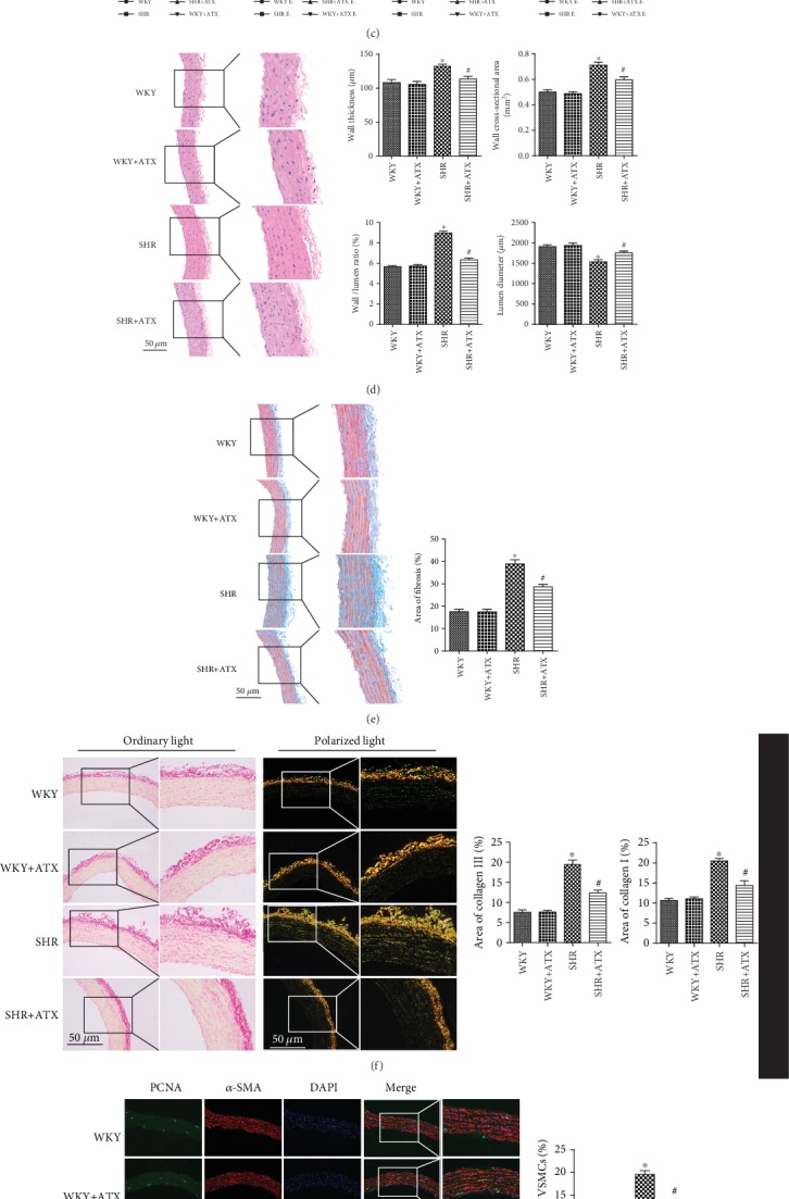
Oxidative stress aggravates mitochondrial injuries and accelerates the proliferation of vascular smooth muscle cells (VSMCs), which are important mechanisms contributing to vascular remodeling in hypertension. We put forward the hypothesis that Astaxanthin (ATX), known to possess strong features of antioxidant, could attenuate vascular remodeling by inhibiting VSMC proliferation and improving mitochondrial function. The potential effects of ATX were tested on spontaneously hypertensive rats (SHRs) and cultured VSMCs that injured by angiotensin II (Ang II). The results showed that ATX lowered blood pressure, reduced aortic wall thickness and fibrosis, and decreased the level of reactive oxygen species (ROS) and HO in tunica media. Moreover, ATX decreased the expression of proliferating cell nuclear antigen (PCNA) and ki67 in aortic VSMCs. , ATX mitigated VSMC proliferation and migration, decreased the level of cellular ROS, and balanced the activities of ROS-related enzymes including NADPH oxidase, xanthine oxidase, and superoxide dismutase (SOD). Besides, ATX mitigated Ca overload, the overproduction of mitochondrial ROS (mtROS), mitochondrial dysfunction, mitochondrial fission, and Drp1 phosphorylation at Ser616. In addition, ATX enhanced mitophagy and mitochondrial biosynthesis by increasing the expression of PINK, parkin, mtDNA, mitochondrial transcription factor A (Tfam), and PGC-1. The present study indicated that ATX could efficiently treat vascular remodeling through restraining VSMC proliferation and restoring mitochondrial function. Inhibiting mitochondrial fission by decreasing the phosphorylation of Drp1 and stimulating mitochondrial autophagy and biosynthesis via increasing the expression of PINK, parkin, Tfam, and PGC-1 may be part of its underlying mechanisms.
氧化应激加剧线粒体损伤并加速血管平滑肌细胞(VSMCs)的增殖,这是高血压血管重构的重要机制。我们提出假设,虾青素(ATX)具有很强的抗氧化特性,可通过抑制 VSMC 增殖和改善线粒体功能来减轻血管重构。在自发性高血压大鼠(SHRs)和血管紧张素 II(Ang II)损伤的培养 VSMCs 上测试了 ATX 的潜在作用。结果表明,ATX 降低血压,降低主动脉壁厚度和纤维化,并降低中层中的活性氧(ROS)和 HO 水平。此外,ATX 降低了主动脉 VSMCs 中增殖细胞核抗原(PCNA)和 ki67 的表达。ATX 减轻了 VSMC 的增殖和迁移,降低了细胞 ROS 水平,并平衡了包括 NADPH 氧化酶、黄嘌呤氧化酶和超氧化物歧化酶(SOD)在内的 ROS 相关酶的活性。此外,ATX 通过减轻 Ca 超载、线粒体 ROS(mtROS)的过度产生、线粒体功能障碍、线粒体分裂和 Drp1 在 Ser616 处的磷酸化来减轻 Ca 超载。此外,ATX 通过增加 PINK、parkin、mtDNA、线粒体转录因子 A(Tfam)和 PGC-1 的表达来增强线粒体自噬和生物合成。本研究表明,ATX 通过抑制 VSMC 增殖和恢复线粒体功能来有效治疗血管重构。通过降低 Drp1 的磷酸化并通过增加 PINK、parkin、Tfam 和 PGC-1 的表达来刺激线粒体自噬和生物合成来抑制线粒体分裂可能是其潜在机制的一部分。