Diaz-Galvan Patricia, Miyagawa Toji, Przybelski Scott A, Lesnick Timothy G, Senjem Matthew L, Jack Clifford R, Forsberg Leah K, Min Hoon-Ki, St Louis Erik K, Savica Rodolfo, Fields Julie A, Benarroch Eduardo E, Lowe Val, Petersen Ronald C, Boeve Bradley F, Kantarci Kejal
Department of Radiology, Mayo Clinic, Rochester, MN 55905, USA.
Department of Neurology, Mayo Clinic, Rochester, MN 55905, USA.
Brain Commun. 2023 Feb 2;5(1):fcad021. doi: 10.1093/braincomms/fcad021. eCollection 2023.
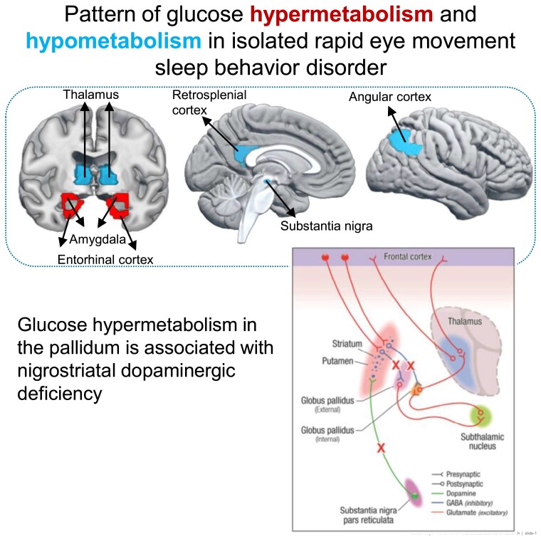
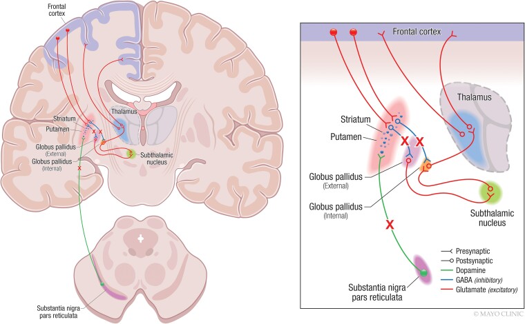
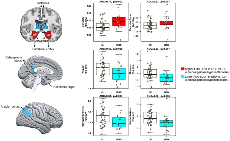
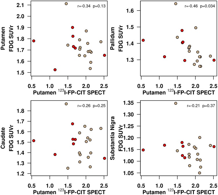
Alterations of cerebral glucose metabolism can be detected in patients with isolated rapid eye movement sleep behaviour disorder, a prodromal feature of neurodegenerative diseases with α-synuclein pathology. However, metabolic characteristics that determine clinical progression in isolated rapid eye movement sleep behaviour disorder and their association with other biomarkers need to be elucidated. We investigated the pattern of cerebral glucose metabolism on F-fluorodeoxyglucose PET in patients with isolated rapid eye movement sleep behaviour disorder, differentiating between those who clinically progressed and those who remained stable over time. Second, we studied the association between F-fluorodeoxyglucose PET and lower dopamine transporter availability in the putamen, another hallmark of synucleinopathies. Patients with isolated rapid eye movement sleep behaviour disorder from the Mayo Clinic Alzheimer's Disease Research Center and Center for Sleep Medicine ( = 22) and age-and sex-matched clinically unimpaired controls (clinically unimpaired; = 44) from the Mayo Clinic Study of Aging were included. All participants underwent F-fluorodeoxyglucose PET and dopamine transporter imaging with iodine 123-radiolabeled 2β-carbomethoxy-3β-(4-iodophenyl)-N-(3-fluoropropyl) nortropane on single-photon emission computerized tomography. A subset of patients with isolated rapid eye movement sleep behaviour disorder with follow-up evaluations ( = 17) was classified as isolated rapid eye movement sleep behaviour disorder progressors ( = 7) if they developed mild cognitive impairment or Parkinson's disease; or isolated rapid eye movement sleep behaviour disorder stables ( = 10) if they remained with a diagnosis of isolated rapid eye movement sleep behaviour disorder with no cognitive impairment. Glucose metabolic abnormalities in isolated rapid eye movement sleep behaviour disorder were determined by comparing atlas-based regional F-fluorodeoxyglucose PET uptake between isolated rapid eye movement sleep behaviour disorder and clinically unimpaired. Associations between F-fluorodeoxyglucose PET and dopamine transporter availability in the putamen were analyzed with Pearson's correlation within the nigrostriatal pathway structures and with voxel-based analysis in the cortex. Patients with isolated rapid eye movement sleep behaviour disorder had lower glucose metabolism in the substantia nigra, retrosplenial cortex, angular cortex, and thalamus, and higher metabolism in the amygdala and entorhinal cortex compared with clinically unimpaired. Patients with isolated rapid eye movement sleep behaviour disorder who clinically progressed over time were characterized by higher glucose metabolism in the amygdala and entorhinal cortex, and lower glucose metabolism in the cerebellum compared with clinically unimpaired. Lower dopamine transporter availability in the putamen was associated with higher glucose metabolism in the pallidum within the nigrostriatal pathway; and with higher F-fluorodeoxyglucose uptake in the amygdala, insula, and temporal pole on a voxel-based analysis, although these associations did not survive after correcting for multiple comparisons. Our findings suggest that cerebral glucose metabolism in isolated rapid eye movement sleep behaviour disorder is characterized by hypometabolism in regions frequently affected during the prodromal stage of synucleinopathies, potentially reflecting synaptic dysfunction. Hypermetabolism is also seen in isolated rapid eye movement sleep behaviour disorder, suggesting that synaptic metabolic disruptions may be leading to a lack of inhibition, compensatory mechanisms, or microglial activation, especially in regions associated with nigrostriatal degeneration.
在患有孤立性快速眼动睡眠行为障碍的患者中可检测到脑葡萄糖代谢改变,这是一种具有α-突触核蛋白病理特征的神经退行性疾病的前驱特征。然而,需要阐明决定孤立性快速眼动睡眠行为障碍临床进展的代谢特征及其与其他生物标志物的关联。我们研究了孤立性快速眼动睡眠行为障碍患者在F-氟脱氧葡萄糖PET上的脑葡萄糖代谢模式,区分了随时间临床进展的患者和保持稳定的患者。其次,我们研究了F-氟脱氧葡萄糖PET与壳核中多巴胺转运体可用性降低之间的关联,这是突触核蛋白病的另一个标志。纳入了梅奥诊所阿尔茨海默病研究中心和睡眠医学中心的孤立性快速眼动睡眠行为障碍患者(n = 22),以及梅奥诊所衰老研究中年龄和性别匹配的临床无损害对照者(临床无损害;n = 44)。所有参与者均接受了F-氟脱氧葡萄糖PET检查,并在单光子发射计算机断层扫描上使用碘123放射性标记的2β-甲氧基羰基-3β-(4-碘苯基)-N-(3-氟丙基)去甲托烷进行多巴胺转运体成像。对一部分有随访评估的孤立性快速眼动睡眠行为障碍患者(n = 17)进行分类,如果他们发展为轻度认知障碍或帕金森病,则分类为孤立性快速眼动睡眠行为障碍进展者(n = 7);如果他们仍被诊断为孤立性快速眼动睡眠行为障碍且无认知障碍,则分类为孤立性快速眼动睡眠行为障碍稳定者(n = 10)。通过比较孤立性快速眼动睡眠行为障碍患者与临床无损害者基于图谱的区域F-氟脱氧葡萄糖PET摄取情况,确定孤立性快速眼动睡眠行为障碍中的葡萄糖代谢异常。在黑质纹状体通路结构内,使用Pearson相关性分析F-氟脱氧葡萄糖PET与壳核中多巴胺转运体可用性之间的关联,并在皮质中进行基于体素的分析。与临床无损害者相比,孤立性快速眼动睡眠行为障碍患者在黑质、扣带回后皮质、角回皮质和丘脑中的葡萄糖代谢较低,而在杏仁核和内嗅皮质中的代谢较高。随时间临床进展的孤立性快速眼动睡眠行为障碍患者的特征是,与临床无损害者相比,杏仁核和内嗅皮质中的葡萄糖代谢较高,而小脑中的葡萄糖代谢较低。壳核中多巴胺转运体可用性降低与黑质纹状体通路内苍白球中的葡萄糖代谢较高相关;在基于体素的分析中,与杏仁核、岛叶和颞极中较高的F-氟脱氧葡萄糖摄取相关,尽管在进行多重比较校正后这些关联不再显著。我们的研究结果表明,孤立性快速眼动睡眠行为障碍中的脑葡萄糖代谢特征是在突触核蛋白病前驱期经常受影响的区域存在代谢减低,这可能反映了突触功能障碍。在孤立性快速眼动睡眠行为障碍中也可见代谢增高,这表明突触代谢紊乱可能导致抑制缺乏、代偿机制或小胶质细胞激活,特别是在与黑质纹状体变性相关的区域。