Department of Nuclear Medicine, State Key Laboratory of Complex Severe and Rare Diseases, Beijing Key Laboratory of Molecular Targeted Diagnosis and Therapy in Nuclear Medicine, Peking Union Medical College Hospital, Chinese Academy of Medical Sciences, Beijing, 100730, China.
Medical Science Research Center (MRC), Peking Union Medical College Hospital, Chinese Academy of Medical Sciences, Beijing, 100730, China.
BMC Med Imaging. 2023 Feb 27;23(1):35. doi: 10.1186/s12880-023-00987-7.
The maximum likelihood activity and attenuation (MLAA) reconstruction algorithm has been proposed to jointly estimate tracer activity and attenuation at the same time, and proven to be a promising solution to the CT attenuation correction (CT-AC) artifacts in PET images. This study aimed to perform a quantitative evaluation and clinical validation of the MLAA method.
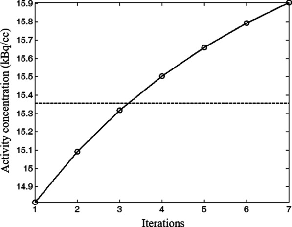
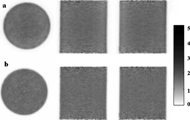
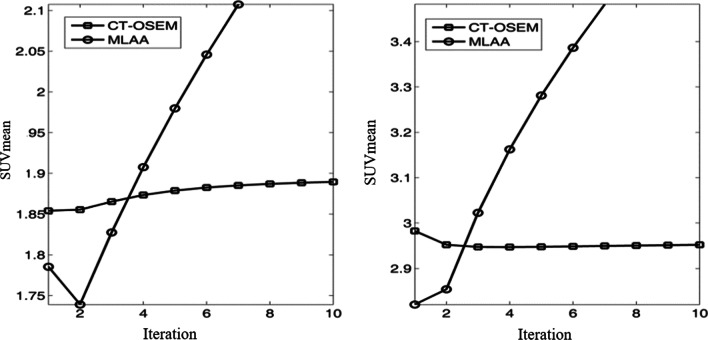
A uniform cylinder phantom filled with F-FDG solution was scanned to optimize the reconstruction parameters for the implemented MLAA algorithm. 67 patients who underwent whole-body F-FDG PET/CT scan were retrospectively recruited. PET images were reconstructed using MLAA and clinical standard OSEM algorithm with CT-AC (CT-OSEM). The mean and maximum standardized uptake values (SUVmean and SUVmax) in regions of interest (ROIs) of organs, high uptake lesions and areas affected by metal implants and respiration motion artifacts were quantitatively analyzed.
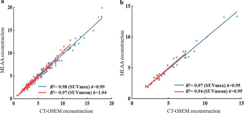
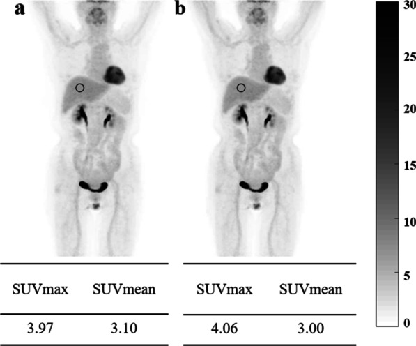
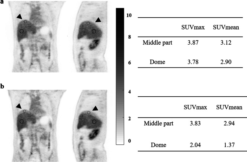
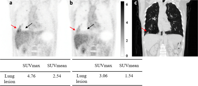
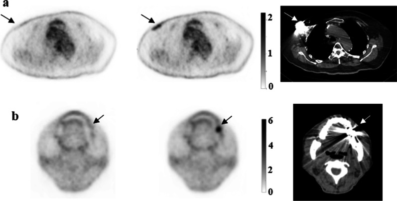
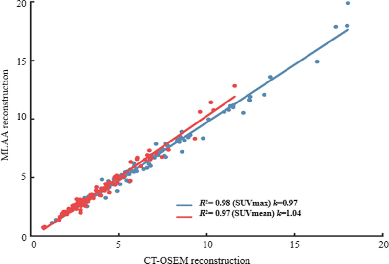
In quantitative analysis, SUVs in patient's organ ROIs between two methods showed R ranging from 0.91 to 0.98 and k ranging from 0.90 to 1.06, and the average SUVmax and SUVmean differences between two methods were within 10% range, except for the lung ROI, which was 10.5% and 16.73% respectively. The average SUVmax and SUVmean differences of a total of 117 high uptake lesions were 7.25% and 7.10% respectively. 20 patients were identified to have apparent respiration motion artifacts in the liver in CT-OSEM images, and the SUVs differences between two methods measured at dome of the liver were significantly larger than measured at middle part of the liver. 10 regions with obvious metal artifacts were identified in CT-OSEM images and the average SUVmean and SUVmax differences in metal implants affected regions were reported to be 52.90% and 56.20% respectively.
PET images reconstructed using MLAA are clinically acceptable in terms of image quality as well as quantification and it is a useful tool in clinical practice, especially when CT-AC may cause respiration motion and metal artifacts. Moreover, this study also provides technical reference and data support for the future iteration and development of PET reconstruction technology of SUV accurate quantification.
最大似然活性和衰减(MLAA)重建算法已被提出,以同时估计示踪剂活性和衰减,并已被证明是解决 PET 图像中 CT 衰减校正(CT-AC)伪影的有前途的解决方案。本研究旨在对 MLAA 方法进行定量评估和临床验证。
使用充满 F-FDG 溶液的均匀圆柱体体模扫描,以优化实现的 MLAA 算法的重建参数。回顾性招募了 67 名接受全身 F-FDG PET/CT 扫描的患者。使用 MLAA 和具有 CT-AC(CT-OSEM)的临床标准 OSEM 算法重建 PET 图像。定量分析器官、高摄取病变以及受金属植入物和呼吸运动伪影影响的区域的感兴趣区域(ROI)中的平均和最大标准化摄取值(SUVmean 和 SUVmax)。
在定量分析中,两种方法在患者器官 ROI 中的 SUV 之间的 R 值范围为 0.91 到 0.98,k 值范围为 0.90 到 1.06,并且两种方法之间的 SUVmax 和 SUVmean 差异均在 10%范围内,除了肺 ROI,分别为 10.5%和 16.73%。总共 117 个高摄取病变的 SUVmax 和 SUVmean 差异分别为 7.25%和 7.10%。在 CT-OSEM 图像中,20 名患者的肝脏有明显的呼吸运动伪影,并且在肝脏穹顶处测量的两种方法之间的 SUV 差异明显大于在肝脏中部测量的 SUV 差异。在 CT-OSEM 图像中确定了 10 个有明显金属伪影的区域,并且受金属植入物影响区域的 SUVmean 和 SUVmax 差异分别报告为 52.90%和 56.20%。
从图像质量和定量方面来看,使用 MLAA 重建的 PET 图像在临床应用中是可以接受的,它是一种在临床实践中的有用工具,特别是当 CT-AC 可能导致呼吸运动和金属伪影时。此外,本研究还为未来的 SUV 精确定量 PET 重建技术的迭代和发展提供了技术参考和数据支持。