Department of Pharmacology and Vanderbilt Brain Institute, Vanderbilt University, Nashville, TN, USA.
STAR Protoc. 2023 Mar 17;4(1):102080. doi: 10.1016/j.xpro.2023.102080. Epub 2023 Feb 3.
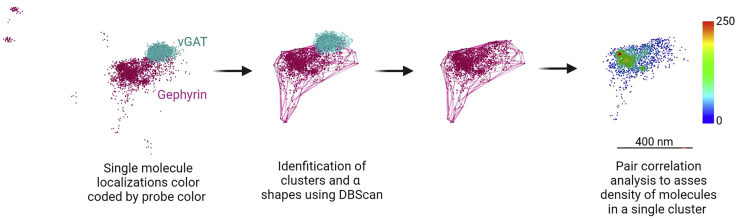
Visualizing the nano-organization of the synapse is fundamental to elucidating the structure-function relationship of the nervous system. The advent of super-resolution microscopy provides a tool to assess and quantify the dynamic organization of numerous proteins at the synapse. Here we present a protocol assessing inhibitory synapse scaffold protein, gephyrin, in rat primary hippocampal cultures using dSTORM microscopy. We delineate the steps for artemisinin treatment, immunocytochemistry, dSTORM image acquisition, single-molecule localization, and the analysis of synaptic scaffold protein dynamics. For complete details on the use and execution of this protocol, please refer to Guzikowski and Kavalali (2022)..
可视化突触的纳米组织对于阐明神经系统的结构-功能关系至关重要。超分辨率显微镜的出现为评估和量化突触中众多蛋白质的动态组织提供了一种工具。在这里,我们使用 dSTORM 显微镜展示了一种评估大鼠原代海马培养物中抑制性突触支架蛋白胶质纤维酸性蛋白 (gephyrin) 的方案。我们描述了青蒿素处理、免疫细胞化学、dSTORM 图像采集、单分子定位和突触支架蛋白动力学分析的步骤。有关此方案使用和执行的完整详细信息,请参阅 Guzikowski 和 Kavalali (2022)。