Yam Patricia T, Pincus Zachary, Gupta Gagan D, Bashkurov Mikhail, Charron Frédéric, Pelletier Laurence, Colman David R
Department of Neurology and Neurosurgery, Montreal Neurological Institute and Hospital, McGill University, Montreal, Quebec, Canada ; Program in Neuroengineering, McGill University, Montreal, Quebec, Canada.
PLoS One. 2013 Nov 1;8(11):e79679. doi: 10.1371/journal.pone.0079679. eCollection 2013.
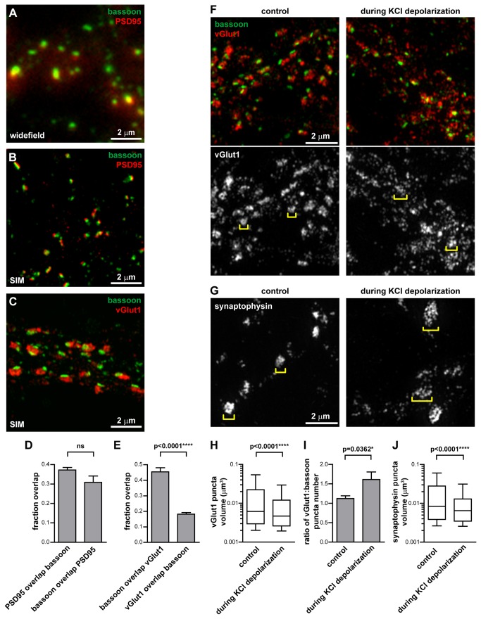
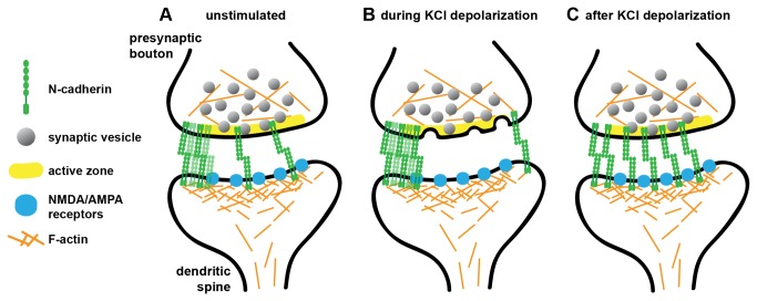
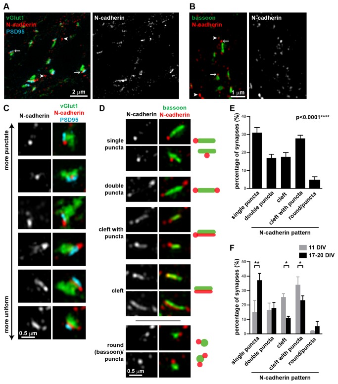
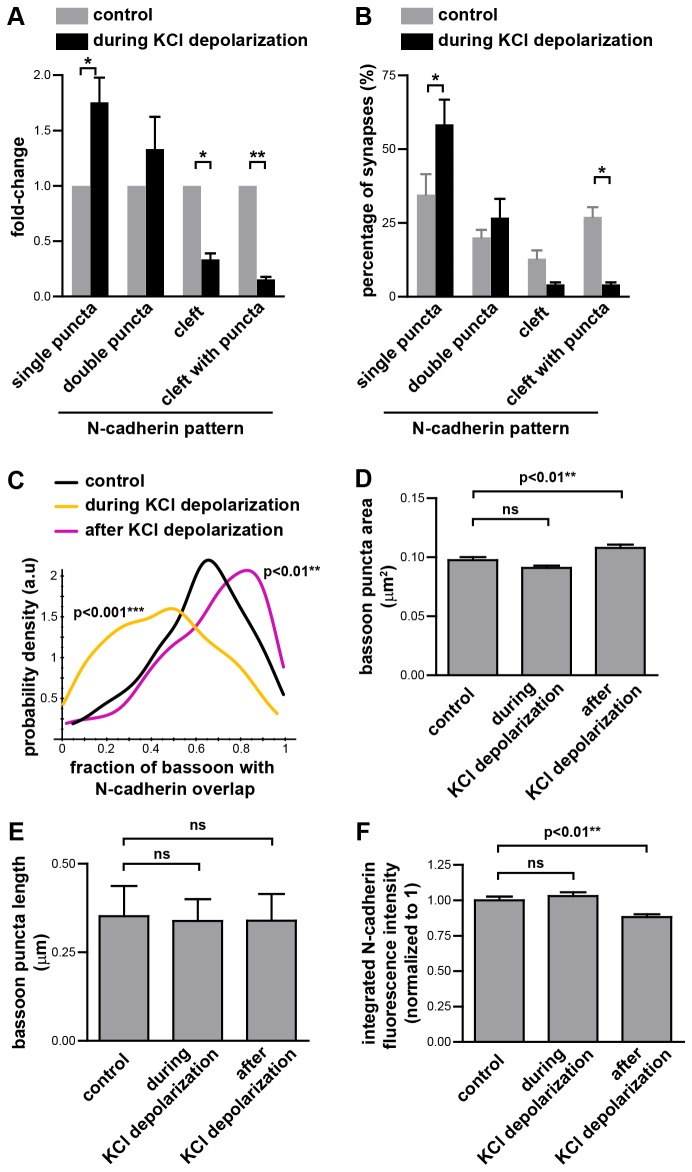
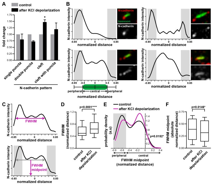
N-cadherin is a cell adhesion molecule which is enriched at synapses. Binding of N-cadherin molecules to each other across the synaptic cleft has been postulated to stabilize adhesion between the presynaptic bouton and the postsynaptic terminal. N-cadherin is also required for activity-induced changes at synapses, including hippocampal long term potentiation and activity-induced spine expansion and stabilization. We hypothesized that these activity-dependent changes might involve changes in N-cadherin localization within synapses. To determine whether synaptic activity changes the localization of N-cadherin, we used structured illumination microscopy, a super-resolution approach which overcomes the conventional resolution limits of light microscopy, to visualize the localization of N-cadherin within synapses of hippocampal neurons. We found that synaptic N-cadherin exhibits a spectrum of localization patterns, ranging from puncta at the periphery of the synapse adjacent to the active zone to an even distribution along the synaptic cleft. Furthermore, the N-cadherin localization pattern within synapses changes during KCl depolarization and after transient synaptic stimulation. During KCl depolarization, N-cadherin relocalizes away from the central region of the synaptic cleft to the periphery of the synapse. In contrast, after transient synaptic stimulation with KCl followed by a period of rest in normal media, fewer synapses have N-cadherin present as puncta at the periphery and more synapses have N-cadherin present more centrally and uniformly along the synapse compared to unstimulated cells. This indicates that transient synaptic stimulation modulates N-cadherin localization within the synapse. These results bring new information to the structural organization and activity-induced changes occurring at synapses, and suggest that N-cadherin relocalization may contribute to activity dependent changes at synapses.
N-钙黏蛋白是一种在突触处富集的细胞黏附分子。据推测,N-钙黏蛋白分子在突触间隙相互结合可稳定突触前终扣与突触后终末之间的黏附。突触处活动诱导的变化,包括海马体长期增强以及活动诱导的树突棘扩张和稳定,也需要N-钙黏蛋白。我们推测,这些依赖于活动的变化可能涉及突触内N-钙黏蛋白定位的改变。为了确定突触活动是否会改变N-钙黏蛋白的定位,我们使用了结构照明显微镜,这是一种超分辨率方法,克服了传统光学显微镜的分辨率限制,以可视化海马神经元突触内N-钙黏蛋白的定位。我们发现,突触N-钙黏蛋白呈现出一系列定位模式,从突触周边靠近活性区的点状分布到沿突触间隙的均匀分布。此外,在氯化钾去极化期间以及短暂突触刺激后,突触内N-钙黏蛋白的定位模式会发生变化。在氯化钾去极化期间,N-钙黏蛋白从突触间隙的中央区域重新定位到突触周边。相比之下,在用氯化钾进行短暂突触刺激后,在正常培养基中休息一段时间,与未受刺激的细胞相比,较少突触的周边有作为点状分布的N-钙黏蛋白,而更多突触的N-钙黏蛋白在突触中央且沿突触更均匀地分布。这表明短暂突触刺激可调节突触内N-钙黏蛋白的定位。这些结果为突触处发生的结构组织和活动诱导的变化带来了新信息,并表明N-钙黏蛋白的重新定位可能有助于突触处依赖于活动的变化。