• 文献检索
  • 文档翻译
  • 深度研究
  • 学术资讯
  • Suppr Zotero 插件Zotero 插件
  • 邀请有礼
  • 套餐&价格
  • 历史记录
应用&插件
Suppr Zotero 插件Zotero 插件浏览器插件Mac 客户端Windows 客户端微信小程序
定价
高级版会员购买积分包购买API积分包
服务
文献检索文档翻译深度研究API 文档MCP 服务
关于我们
关于 Suppr公司介绍联系我们用户协议隐私条款
关注我们

Suppr 超能文献

核心技术专利:CN118964589B侵权必究
粤ICP备2023148730 号-1Suppr @ 2026

文献检索

告别复杂PubMed语法,用中文像聊天一样搜索,搜遍4000万医学文献。AI智能推荐,让科研检索更轻松。

立即免费搜索

文件翻译

保留排版,准确专业,支持PDF/Word/PPT等文件格式,支持 12+语言互译。

免费翻译文档

深度研究

AI帮你快速写综述,25分钟生成高质量综述,智能提取关键信息,辅助科研写作。

立即免费体验

三位同胞兄妹的临床病理发现:地图状萎缩。

Clinicopathologic Findings in Three Siblings With Geographic Atrophy.

机构信息

Wilmer Eye Institute, Johns Hopkins Hospital, Baltimore, Maryland, United States.

Department of Ophthalmology, Bascom Palmer Eye Institute, University of Miami Miller School of Medicine, Miami, Florida, United States.

出版信息

Invest Ophthalmol Vis Sci. 2023 Mar 1;64(3):2. doi: 10.1167/iovs.64.3.2.

DOI:10.1167/iovs.64.3.2
PMID:36862121
原文链接:https://pmc.ncbi.nlm.nih.gov/articles/PMC9983703/
Abstract

PURPOSE

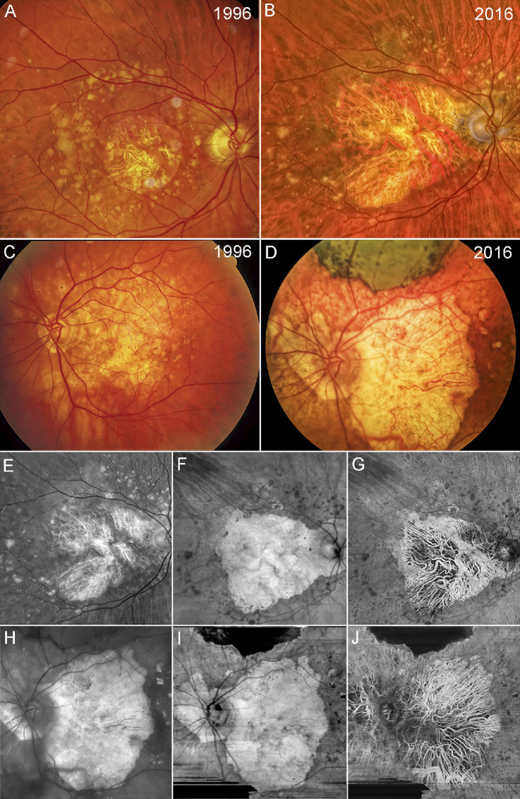
Age-related macular degeneration (AMD) is a leading cause of blindness among the elderly worldwide. Clinical imaging and histopathologic studies are crucial to understanding disease pathology. This study combined clinical observations of three brothers with geographic atrophy (GA), followed for 20 years, with histopathologic analysis.

METHODS

For two of the three brothers, clinical images were taken in 2016, 2 years prior to death. Immunohistochemistry, on both flat-mounts and cross sections, histology, and transmission electron microscopy were used to compare the choroid and retina in GA eyes to those of age-matched controls.

RESULTS

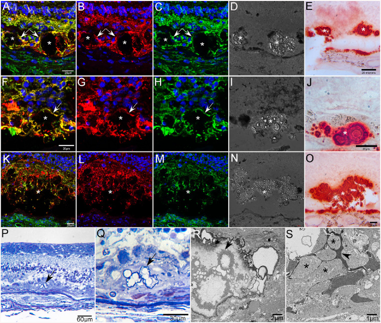
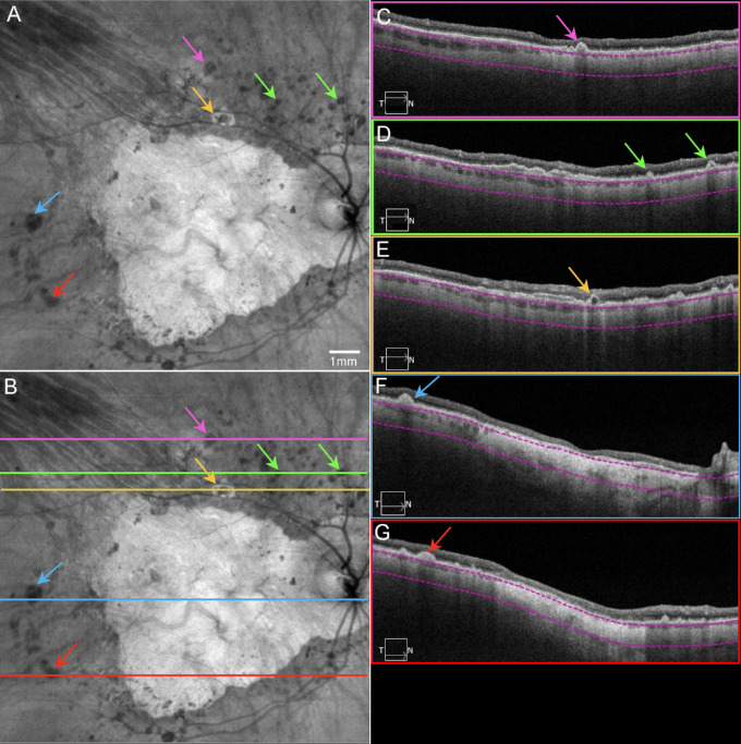
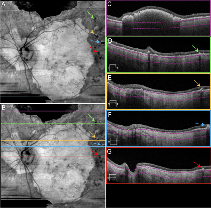
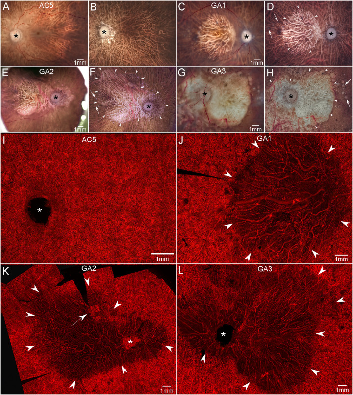
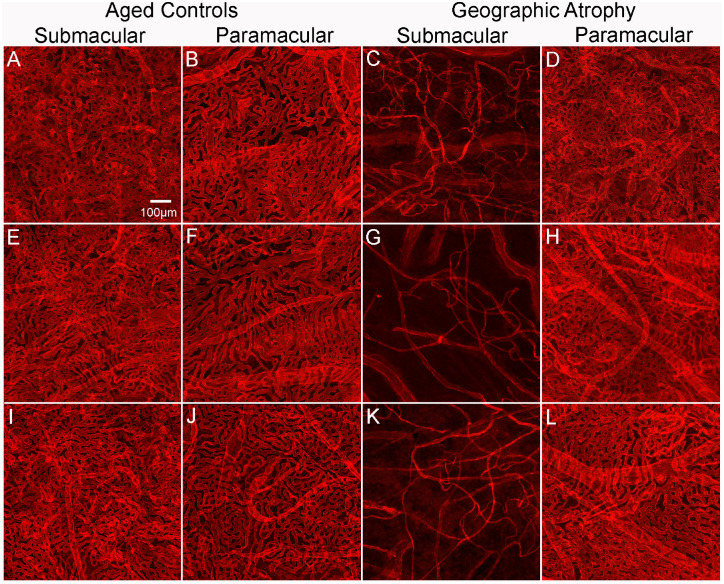
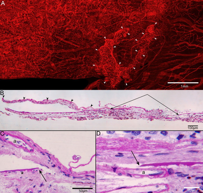
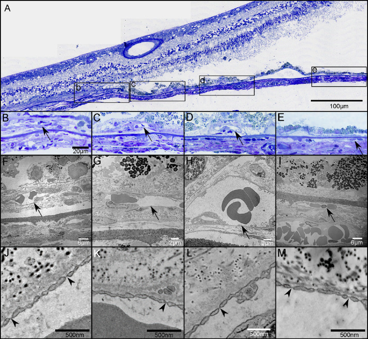
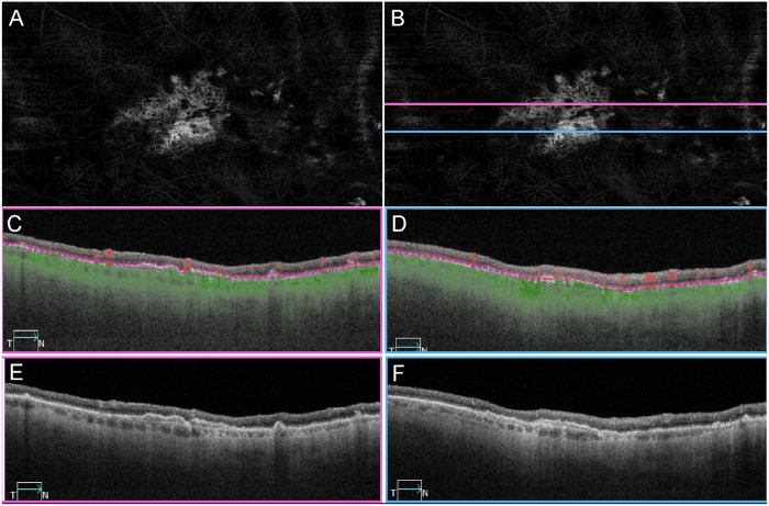
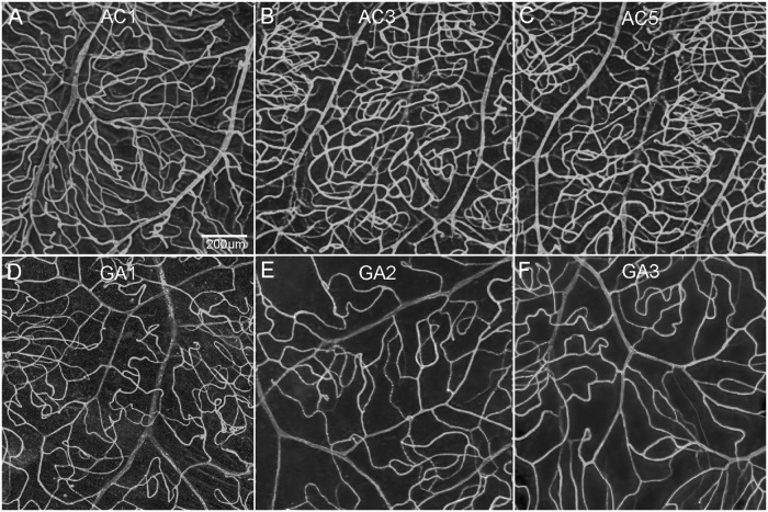
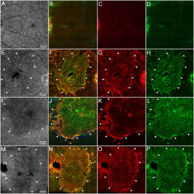
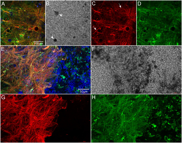
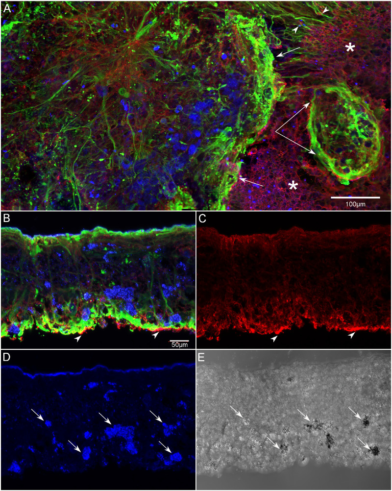
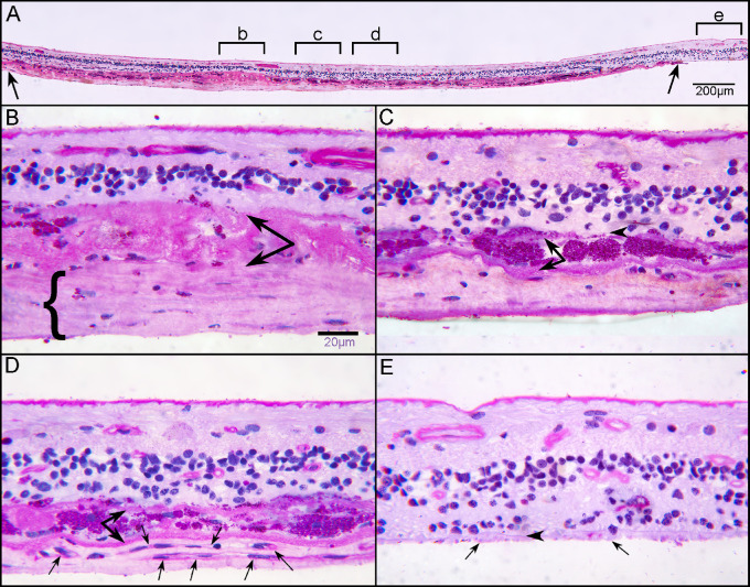
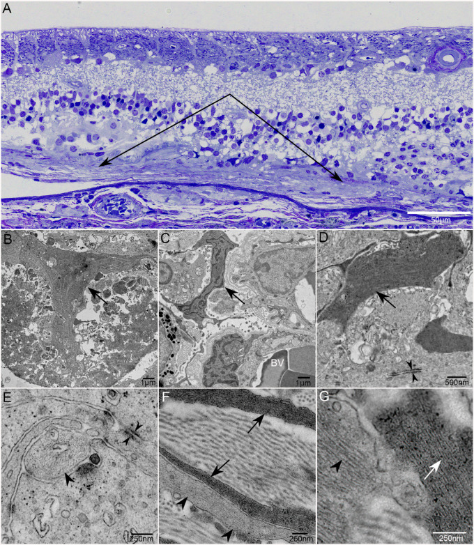
Ulex europaeus agglutinin (UEA) lectin staining of the choroid demonstrated a significant reduction in the percent vascular area and vessel diameter. In one donor, histopathologic analysis demonstrated two separate areas with choroidal neovascularization (CNV). Reevaluation of swept-source optical coherence tomography angiography (SS-OCTA) images revealed CNV in two of the brothers. UEA lectin also revealed a significant reduction in retinal vasculature in the atrophic area. A subretinal glial membrane, composed of processes positive for glial fibrillary acidic protein and/or vimentin, occupied areas identical to those of retinal pigment epithelium (RPE) and choroidal atrophy in all three AMD donors. SS-OCTA also demonstrated presumed calcific drusen in the two donors imaged in 2016. Immunohistochemical analysis and alizarin red S staining verified calcium within drusen, which was ensheathed by glial processes.

CONCLUSIONS

This study demonstrates the importance of clinicohistopathologic correlation studies. It emphasizes the need to better understand how the symbiotic relationship between choriocapillaris and RPE, glial response, and calcified drusen impact GA progression.

摘要

目的

年龄相关性黄斑变性(AMD)是全球老年人致盲的主要原因。临床成像和组织病理学研究对于了解疾病病理学至关重要。本研究结合了三名患有地图状萎缩(GA)的兄弟的临床观察结果,这些兄弟经过 20 年的随访,并结合了组织病理学分析。

方法

对于其中的两名兄弟,在他们去世前两年的 2016 年拍摄了临床图像。使用免疫组织化学、平展和切片组织学以及透射电子显微镜,比较 GA 眼的脉络膜和视网膜与年龄匹配的对照组。

结果

Ulex europaeus agglutinin(UEA)凝集素对脉络膜的染色显示血管面积和血管直径的百分比显著减少。在一名供体中,组织病理学分析显示存在两个独立的脉络膜新生血管化(CNV)区域。重新评估扫频源光学相干断层扫描血管造影(SS-OCTA)图像显示,两名兄弟中有两名存在 CNV。UEA 凝集素也显示出萎缩区域视网膜血管的显著减少。由胶质纤维酸性蛋白和/或波形蛋白阳性的过程组成的视网膜下胶质膜,占据了与所有三名 AMD 供体的 RPE 和脉络膜萎缩相同的区域。SS-OCTA 还显示了在 2016 年成像的两名供体中存在推测的钙化玻璃膜疣。免疫组织化学分析和茜素红 S 染色证实了玻璃膜疣内的钙,钙被胶质过程包裹。

结论

本研究证明了临床组织病理学相关性研究的重要性。它强调了需要更好地了解脉络膜毛细血管和 RPE 之间的共生关系、胶质反应以及钙化玻璃膜疣如何影响 GA 的进展。

https://cdn.ncbi.nlm.nih.gov/pmc/blobs/f04b/9983703/40509923fc3a/iovs-64-3-2-f015.jpg
https://cdn.ncbi.nlm.nih.gov/pmc/blobs/f04b/9983703/87d14d142ce0/iovs-64-3-2-f001.jpg
https://cdn.ncbi.nlm.nih.gov/pmc/blobs/f04b/9983703/913e696070dc/iovs-64-3-2-f002.jpg
https://cdn.ncbi.nlm.nih.gov/pmc/blobs/f04b/9983703/85e3633ade7a/iovs-64-3-2-f003.jpg
https://cdn.ncbi.nlm.nih.gov/pmc/blobs/f04b/9983703/cce87b1aae1a/iovs-64-3-2-f004.jpg
https://cdn.ncbi.nlm.nih.gov/pmc/blobs/f04b/9983703/aa7bdd0d918e/iovs-64-3-2-f005.jpg
https://cdn.ncbi.nlm.nih.gov/pmc/blobs/f04b/9983703/abb9ae31d095/iovs-64-3-2-f006.jpg
https://cdn.ncbi.nlm.nih.gov/pmc/blobs/f04b/9983703/bcc0fb79161d/iovs-64-3-2-f007.jpg
https://cdn.ncbi.nlm.nih.gov/pmc/blobs/f04b/9983703/2a7a841abb9c/iovs-64-3-2-f008.jpg
https://cdn.ncbi.nlm.nih.gov/pmc/blobs/f04b/9983703/fea573036d2d/iovs-64-3-2-f009.jpg
https://cdn.ncbi.nlm.nih.gov/pmc/blobs/f04b/9983703/c49aa5ac001b/iovs-64-3-2-f010.jpg
https://cdn.ncbi.nlm.nih.gov/pmc/blobs/f04b/9983703/a98ffd281477/iovs-64-3-2-f011.jpg
https://cdn.ncbi.nlm.nih.gov/pmc/blobs/f04b/9983703/bcf005c19661/iovs-64-3-2-f012.jpg
https://cdn.ncbi.nlm.nih.gov/pmc/blobs/f04b/9983703/86a5d2ea71a9/iovs-64-3-2-f013.jpg
https://cdn.ncbi.nlm.nih.gov/pmc/blobs/f04b/9983703/db8e3b89f32e/iovs-64-3-2-f014.jpg
https://cdn.ncbi.nlm.nih.gov/pmc/blobs/f04b/9983703/40509923fc3a/iovs-64-3-2-f015.jpg

相似文献

1
Clinicopathologic Findings in Three Siblings With Geographic Atrophy.三位同胞兄妹的临床病理发现:地图状萎缩。
Invest Ophthalmol Vis Sci. 2023 Mar 1;64(3):2. doi: 10.1167/iovs.64.3.2.
2
Histopathological Insights Into Choroidal Vascular Loss in Clinically Documented Cases of Age-Related Macular Degeneration.对临床记录的年龄相关性黄斑变性病例中脉络膜血管丧失的组织病理学见解
JAMA Ophthalmol. 2016 Nov 1;134(11):1272-1280. doi: 10.1001/jamaophthalmol.2016.3519.
3
Optical Coherence Tomography Reflective Drusen Substructures Predict Progression to Geographic Atrophy in Age-related Macular Degeneration.光学相干断层扫描反射性玻璃膜疣亚结构预测年龄相关性黄斑变性进展为地图状萎缩
Ophthalmology. 2016 Dec;123(12):2554-2570. doi: 10.1016/j.ophtha.2016.08.047. Epub 2016 Oct 25.
4
Choriocapillaris dropout in early age-related macular degeneration.脉络膜毛细血管萎缩在早发性年龄相关性黄斑变性中的作用。
Exp Eye Res. 2020 Mar;192:107939. doi: 10.1016/j.exer.2020.107939. Epub 2020 Jan 24.
5
Subretinal Glial Membranes in Eyes With Geographic Atrophy.地图样萎缩眼中的视网膜下胶质膜
Invest Ophthalmol Vis Sci. 2017 Mar 1;58(3):1352-1367. doi: 10.1167/iovs.16-21229.
6
Quantifying changes in RPE and choroidal vasculature in eyes with age-related macular degeneration.量化年龄相关性黄斑变性患者眼睛中视网膜色素上皮和脉络膜血管系统的变化。
Invest Ophthalmol Vis Sci. 2002 Jun;43(6):1986-93.
7
Optical Coherence Tomography Features Preceding the Onset of Advanced Age-Related Macular Degeneration.年龄相关性黄斑变性晚期发病前的光学相干断层扫描特征
Invest Ophthalmol Vis Sci. 2017 Jul 1;58(9):3519-3529. doi: 10.1167/iovs.17-21696.
8
Impact of Drusen Burden on Incidence of Subclinical CNV With OCTA.基于光学相干断层扫描血管造影(OCTA)的玻璃膜疣负荷对亚临床脉络膜新生血管发生率的影响
Ophthalmic Surg Lasers Imaging Retina. 2019 Dec 1;51(1):22-30. doi: 10.3928/23258160-20191211-03.
9
Retinal Glial and Choroidal Vascular Pathology in Donors Clinically Diagnosed With Stargardt Disease.临床诊断为 Stargardt 病的供体的视网膜胶质和脉络膜血管病理学。
Invest Ophthalmol Vis Sci. 2020 Jul 1;61(8):27. doi: 10.1167/iovs.61.8.27.
10
Histologic and morphometric analysis of the choroid, Bruch's membrane, and retinal pigment epithelium in postmortem eyes with age-related macular degeneration and histologic examination of surgically excised choroidal neovascular membranes.对患有年龄相关性黄斑变性的死后眼球的脉络膜、布鲁赫膜和视网膜色素上皮进行组织学和形态计量学分析,并对手术切除的脉络膜新生血管膜进行组织学检查。
Surv Ophthalmol. 1999 Oct;44 Suppl 1:S10-32. doi: 10.1016/s0039-6257(99)00086-7.

引用本文的文献

1
Retinal Vessel Changes in Geographic Atrophy in AMD: Insights From Imaging and Histology.年龄相关性黄斑变性地图样萎缩中的视网膜血管变化:来自影像学和组织学的见解
Invest Ophthalmol Vis Sci. 2025 Aug 1;66(11):69. doi: 10.1167/iovs.66.11.69.
2
Neuroprotection provided by polyphenols and flavonoids in photoreceptor degenerative diseases.多酚和黄酮类化合物在光感受器退行性疾病中的神经保护作用。
Neural Regen Res. 2026 Mar 1;21(3):908-922. doi: 10.4103/NRR.NRR-D-24-01638. Epub 2025 May 6.
3
Dicer Loss in Müller Glia Leads to a Defined Sequence of Pathological Events Beginning With Cone Dysfunction.

本文引用的文献

1
Glial remodeling and choroidal vascular pathology in eyes from two donors with Choroideremia.两名脉络膜视网膜萎缩症供体眼睛中的神经胶质重塑和脉络膜血管病变
Front Ophthalmol (Lausanne). 2022 Oct 14;2:994566. doi: 10.3389/fopht.2022.994566. eCollection 2022.
2
CHANGE IN CHORIOCAPILLARIS FLOW DEFICITS WITHIN TEARS OF THE RETINAL PIGMENT EPITHELIUM IMAGED WITH SWEPT-SOURCE OPTICAL COHERENCE TOMOGRAPHY ANGIOGRAPHY.应用扫频源光学相干断层扫描血管造影术观察到视网膜色素上皮层撕裂处脉络膜血流缺损的变化。
Retina. 2022 Nov 1;42(11):2031-2038. doi: 10.1097/IAE.0000000000003577.
3
Towards a better understanding of non-exudative choroidal and macular neovascularization.
缪勒胶质细胞中Dicer缺失导致一系列特定的病理事件,始于视锥细胞功能障碍。
Invest Ophthalmol Vis Sci. 2025 Mar 3;66(3):7. doi: 10.1167/iovs.66.3.7.
4
Capturing the transition from intermediate to neovascular AMD: Longitudinal changes in choroidal volume and choroidal vascularity index.捕捉中间型到新生血管性年龄相关性黄斑变性的转变:脉络膜体积和脉络膜血管指数的纵向变化。
Graefes Arch Clin Exp Ophthalmol. 2025 Feb 13. doi: 10.1007/s00417-025-06767-z.
5
Regional Alterations in Müller Cell Protein Expression in Human and a Rat Model of Geographic Atrophy.人类及地图样萎缩大鼠模型中 Müller 细胞蛋白表达的区域改变
Invest Ophthalmol Vis Sci. 2025 Feb 3;66(2):21. doi: 10.1167/iovs.66.2.21.
6
Fundus Autofluorescence Variation in Geographic Atrophy of Age-Related Macular Degeneration: A Clinicopathologic Correlation.年龄相关性黄斑变性地图样萎缩中的眼底自发荧光变化:临床病理相关性研究
Invest Ophthalmol Vis Sci. 2025 Jan 2;66(1):49. doi: 10.1167/iovs.66.1.49.
7
The Umbra and the Penumbra: Longitudinal Effects of Geographic Atrophy in AMD on the Outer Choroid by Imaging Analysis and Histopathological Correlation.本影与半影:通过影像学分析和组织病理学相关性研究年龄相关性黄斑变性中地理萎缩对脉络膜外层的纵向影响
Invest Ophthalmol Vis Sci. 2024 Dec 2;65(14):2. doi: 10.1167/iovs.65.14.2.
8
Choriocapillaris in Age-Related Macular Degeneration.年龄相关性黄斑变性中的脉络膜毛细血管。
Turk J Ophthalmol. 2024 Aug 28;54(4):228-234. doi: 10.4274/tjo.galenos.2024.04608.
9
Altered Elastin Turnover, Immune Response, and Age-Related Retinal Thinning in a Transgenic Mouse Model With RPE-Specific HTRA1 Overexpression.在具有 RPE 特异性 HTRA1 过表达的转基因小鼠模型中,弹性蛋白周转率改变、免疫反应和与年龄相关的视网膜变薄。
Invest Ophthalmol Vis Sci. 2024 Jul 1;65(8):34. doi: 10.1167/iovs.65.8.34.
10
Structural OCT and OCT angiography biomarkers associated with the development and progression of geographic atrophy in AMD.与 AMD 中地图状萎缩的发展和进展相关的结构 OCT 和 OCT 血管造影生物标志物。
Graefes Arch Clin Exp Ophthalmol. 2024 Nov;262(11):3421-3436. doi: 10.1007/s00417-024-06497-8. Epub 2024 Apr 30.
旨在更好地理解非渗出性脉络膜和黄斑新生血管化。
Prog Retin Eye Res. 2023 Jan;92:101113. doi: 10.1016/j.preteyeres.2022.101113. Epub 2022 Aug 13.
4
Multimodal Imaging and OCT Detection of Calcified Drusen in Eyes with Age-Related Macular Degeneration.多模态成像与光学相干断层扫描检测年龄相关性黄斑变性眼中的钙化性玻璃膜疣
Ophthalmol Sci. 2022 Jun;2(2). doi: 10.1016/j.xops.2022.100162. Epub 2022 Apr 20.
5
Local Geographic Atrophy Growth Rates Not Influenced by Close Proximity to Non-Exudative Type 1 Macular Neovascularization.局部地理萎缩增长率不受紧邻非渗出型 1 型黄斑新生血管的影响。
Invest Ophthalmol Vis Sci. 2022 Jan 3;63(1):20. doi: 10.1167/iovs.63.1.20.
6
Persistent Hypertransmission Defects Detected on En Face Swept Source Optical Computed Tomography Images Predict the Formation of Geographic Atrophy in Age-Related Macular Degeneration.在年龄相关性黄斑变性中,基于光学相干断层扫描的实时面扫描源图像上检测到的持续性高传输缺陷可预测地图状萎缩的形成。
Am J Ophthalmol. 2022 May;237:58-70. doi: 10.1016/j.ajo.2021.11.001. Epub 2021 Nov 13.
7
Swept-Source OCT Angiographic Characteristics of Treatment-Naïve Nonexudative Macular Neovascularization in AMD Prior to Exudation.AMD 治疗前未出现渗出的非渗出性黄斑新生血管化的扫频源 OCT 血管造影特征。
Invest Ophthalmol Vis Sci. 2021 May 3;62(6):14. doi: 10.1167/iovs.62.6.14.
8
Age-related macular degeneration.年龄相关性黄斑变性。
Nat Rev Dis Primers. 2021 May 6;7(1):31. doi: 10.1038/s41572-021-00265-2.
9
Correlations Between Choriocapillaris and Choroidal Measurements and the Growth of Geographic Atrophy Using Swept Source OCT Imaging.使用扫频源光学相干断层扫描成像技术测量脉络膜毛细血管和脉络膜与地图样萎缩进展之间的相关性
Am J Ophthalmol. 2021 Apr;224:321-331. doi: 10.1016/j.ajo.2020.12.015. Epub 2020 Dec 24.
10
C5 Inhibitor Avacincaptad Pegol for Geographic Atrophy Due to Age-Related Macular Degeneration: A Randomized Pivotal Phase 2/3 Trial.C5 抑制剂阿伐西普 Pegol 治疗年龄相关性黄斑变性相关地图状萎缩:一项随机关键性 2/3 期试验。
Ophthalmology. 2021 Apr;128(4):576-586. doi: 10.1016/j.ophtha.2020.08.027. Epub 2020 Sep 1.