• 文献检索
  • 文档翻译
  • 深度研究
  • 学术资讯
  • Suppr Zotero 插件Zotero 插件
  • 邀请有礼
  • 套餐&价格
  • 历史记录
应用&插件
Suppr Zotero 插件Zotero 插件浏览器插件Mac 客户端Windows 客户端微信小程序
定价
高级版会员购买积分包购买API积分包
服务
文献检索文档翻译深度研究API 文档MCP 服务
关于我们
关于 Suppr公司介绍联系我们用户协议隐私条款
关注我们

Suppr 超能文献

核心技术专利:CN118964589B侵权必究
粤ICP备2023148730 号-1Suppr @ 2026

文献检索

告别复杂PubMed语法,用中文像聊天一样搜索,搜遍4000万医学文献。AI智能推荐,让科研检索更轻松。

立即免费搜索

文件翻译

保留排版,准确专业,支持PDF/Word/PPT等文件格式,支持 12+语言互译。

免费翻译文档

深度研究

AI帮你快速写综述,25分钟生成高质量综述,智能提取关键信息,辅助科研写作。

立即免费体验

扩散加权成像作为一种定量影像学生物标志物,用于预测肝细胞癌的增殖率:开发放射组学列线图。

Diffusion-Weighted Imaging as a Quantitative Imaging Biomarker for Predicting Proliferation Rate in Hepatocellular Carcinoma: Developing a Radiomics Nomogram.

机构信息

From the Department of Radiology, The First Affiliated Hospital of Fujian Medical University.

出版信息

J Comput Assist Tomogr. 2023;47(4):539-547. doi: 10.1097/RCT.0000000000001448. Epub 2023 Mar 6.

DOI:10.1097/RCT.0000000000001448
PMID:36877762
原文链接:https://pmc.ncbi.nlm.nih.gov/articles/PMC10348622/
Abstract

PURPOSE

This study aimed to explore the predictive performance of diffusion-weighted imaging with apparent diffusion coefficient map in predicting the proliferation rate of hepatocellular carcinoma and to develop a radiomics-based nomogram.

METHODS

This was a single-center retrospective study. A total of 110 patients were enrolled. The sample included 38 patients with low Ki67 expression (Ki67 ≤10%) and 72 with high Ki67 expression (Ki67 >10%) as demonstrated by surgical pathology. Patients were randomly divided into either a training (n = 77) or validation (n = 33) cohort. Diffusion-weighted imaging with apparent diffusion coefficient maps was used to extract radiomic features and the signal intensity values of tumor (SI tumor ), normal liver (SI liver ), and background noise (SI background ) from all samples. Subsequently, the clinical model, radiomic model, and fusion model (with clinical data and radiomic signature) were developed and validated.

RESULTS

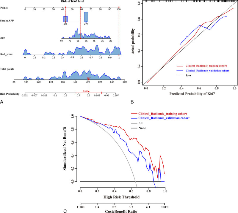
The area under the curve (AUC) of the clinical model for predicting the Ki67 expression including serum α-fetoprotein level ( P = 0.010), age ( P = 0.015), and signal noise ratio ( P = 0.026) was 0.799 and 0.715 in training and validation cohorts, respectively. The AUC of the radiomic model constructed by 9 selected radiomic features was 0.833 and 0.772 in training and validation cohorts, respectively. The AUC of the fusion model containing serum α-fetoprotein level ( P = 0.011), age ( P = 0.019), and rad score ( P < 0.001) was 0.901 and 0.781 in training and validation cohorts, respectively.

CONCLUSIONS

Diffusion-weighted imaging as a quantitative imaging biomarker can predict Ki67 expression level in hepatocellular carcinoma across various models.

摘要

目的

本研究旨在探讨表观扩散系数图弥散加权成像对肝细胞癌增殖率的预测性能,并建立基于放射组学的列线图。

方法

这是一项单中心回顾性研究。共纳入 110 例患者。样本包括 38 例 Ki67 低表达(Ki67≤10%)患者和 72 例 Ki67 高表达(Ki67>10%)患者,均经手术病理证实。患者被随机分为训练集(n=77)和验证集(n=33)。所有样本均采用弥散加权成像表观扩散系数图提取放射组学特征和肿瘤(SI 肿瘤)、正常肝(SI 肝)和背景噪声(SI 背景)的信号强度值。随后,建立并验证了临床模型、放射组学模型和融合模型(结合临床数据和放射组学特征)。

结果

Ki67 表达预测的临床模型(包括血清甲胎蛋白水平(P=0.010)、年龄(P=0.015)和信号噪声比(P=0.026))的曲线下面积(AUC)在训练和验证组中分别为 0.799 和 0.715。由 9 个选定的放射组学特征构建的放射组学模型的 AUC 在训练和验证组中分别为 0.833 和 0.772。包含血清甲胎蛋白水平(P=0.011)、年龄(P=0.019)和 rad 评分(P<0.001)的融合模型的 AUC 在训练和验证组中分别为 0.901 和 0.781。

结论

弥散加权成像作为一种定量成像生物标志物,可以在不同模型中预测肝细胞癌的 Ki67 表达水平。

https://cdn.ncbi.nlm.nih.gov/pmc/blobs/2b7c/10348622/011e3bf79a08/jcat-47-539-g005.jpg
https://cdn.ncbi.nlm.nih.gov/pmc/blobs/2b7c/10348622/e08174300b6f/jcat-47-539-g001.jpg
https://cdn.ncbi.nlm.nih.gov/pmc/blobs/2b7c/10348622/8cd4533aa22c/jcat-47-539-g002.jpg
https://cdn.ncbi.nlm.nih.gov/pmc/blobs/2b7c/10348622/594ea783e968/jcat-47-539-g003.jpg
https://cdn.ncbi.nlm.nih.gov/pmc/blobs/2b7c/10348622/337cc5dffa64/jcat-47-539-g004.jpg
https://cdn.ncbi.nlm.nih.gov/pmc/blobs/2b7c/10348622/011e3bf79a08/jcat-47-539-g005.jpg

相似文献

1
Diffusion-Weighted Imaging as a Quantitative Imaging Biomarker for Predicting Proliferation Rate in Hepatocellular Carcinoma: Developing a Radiomics Nomogram.扩散加权成像作为一种定量影像学生物标志物,用于预测肝细胞癌的增殖率:开发放射组学列线图。
J Comput Assist Tomogr. 2023;47(4):539-547. doi: 10.1097/RCT.0000000000001448. Epub 2023 Mar 6.
2
Radiomics and nomogram of magnetic resonance imaging for preoperative prediction of microvascular invasion in small hepatocellular carcinoma.磁共振成像的放射组学和列线图在小肝细胞癌微血管侵犯术前预测中的应用。
World J Gastroenterol. 2022 Aug 21;28(31):4399-4416. doi: 10.3748/wjg.v28.i31.4399.
3
Prediction of microvascular invasion in hepatocellular carcinoma patients with MRI radiomics based on susceptibility weighted imaging and T2-weighted imaging.基于磁敏感加权成像和 T2 加权成像的 MRI 放射组学预测肝细胞癌患者的微血管侵犯。
Radiol Med. 2024 Aug;129(8):1130-1142. doi: 10.1007/s11547-024-01845-4. Epub 2024 Jul 13.
4
Radiomic analysis of Gd-EOB-DTPA-enhanced MRI predicts Ki-67 expression in hepatocellular carcinoma.钆塞酸二钠增强 MRI 的放射组学分析预测肝细胞癌的 Ki-67 表达。
BMC Med Imaging. 2021 Jun 15;21(1):100. doi: 10.1186/s12880-021-00633-0.
5
A radiomics-based biomarker for cytokeratin 19 status of hepatocellular carcinoma with gadoxetic acid-enhanced MRI.基于影像组学的钆塞酸增强 MRI 肝细胞癌细胞角蛋白 19 状态的生物标志物
Eur Radiol. 2020 May;30(5):3004-3014. doi: 10.1007/s00330-019-06585-y. Epub 2020 Jan 30.
6
Role of the Clinical Features and MRI Parameters on Ki-67 Expression in Hepatocellular Carcinoma Patients: Development of a Predictive Nomogram.肝细胞癌患者临床特征和 MRI 参数对 Ki-67 表达的作用:预测列线图的建立。
J Gastrointest Cancer. 2024 Sep;55(3):1069-1078. doi: 10.1007/s12029-024-01051-5. Epub 2024 Apr 9.
7
Preoperative prediction of macrotrabecular-massive hepatocellular carcinoma through dynamic contrast-enhanced magnetic resonance imaging-based radiomics.基于动态对比增强磁共振成像的放射组学术前预测巨梁型/块状型肝细胞癌。
World J Gastroenterol. 2023 Apr 7;29(13):2001-2014. doi: 10.3748/wjg.v29.i13.2001.
8
Preoperative contrast-enhanced computed tomography-based radiomics model for overall survival prediction in hepatocellular carcinoma.基于术前增强 CT 的影像组学模型预测肝细胞癌患者总生存期。
World J Gastroenterol. 2022 Aug 21;28(31):4376-4389. doi: 10.3748/wjg.v28.i31.4376.
9
A radiomics nomogram for preoperative prediction of microvascular invasion risk in hepatitis B virus-related hepatocellular carcinoma.一种用于术前预测乙型肝炎病毒相关肝细胞癌微血管侵犯风险的影像组学列线图。
Diagn Interv Radiol. 2018 May-Jun;24(3):121-127. doi: 10.5152/dir.2018.17467.
10
Computed tomography radiomic features and clinical factors predicting the response to first transarterial chemoembolization in intermediate-stage hepatocellular carcinoma.基于 CT 影像组学特征与临床因素预测中期肝细胞癌患者首次经动脉化疗栓塞治疗反应的研究
Hepatobiliary Pancreat Dis Int. 2024 Aug;23(4):361-369. doi: 10.1016/j.hbpd.2023.06.011. Epub 2023 Jul 5.

引用本文的文献

1
Radiomics predicting immunohistochemical markers in primary hepatic carcinoma: Current status and challenges.基于影像组学预测原发性肝癌的免疫组化标志物:现状与挑战
Heliyon. 2024 Nov 20;10(23):e40588. doi: 10.1016/j.heliyon.2024.e40588. eCollection 2024 Dec 15.
2
Prognostic value of FDX1, the cuprotosis key gene, and its prediction models across imaging modalities and histology.FDX1(铜死亡关键基因)的预后价值及其在影像学和组织学上的预测模型。
BMC Cancer. 2024 Nov 11;24(1):1381. doi: 10.1186/s12885-024-13149-x.
3
Leveraging radiomics and AI for precision diagnosis and prognostication of liver malignancies.

本文引用的文献

1
Correlationship between Ki67, VEGF, and p53 and Hepatocellular Carcinoma Recurrence in Liver Transplant Patients.Ki67、VEGF 和 p53 与肝移植患者肝癌复发的相关性。
Biomed Res Int. 2021 Apr 15;2021:6651397. doi: 10.1155/2021/6651397. eCollection 2021.
2
Evaluation of the Therapeutic Effect of Adjuvant Transcatheter Arterial Chemoembolization Based on Ki67 After Hepatocellular Carcinoma Surgery.基于Ki67评估肝细胞癌术后辅助经动脉化疗栓塞的治疗效果
Front Oncol. 2021 Feb 25;11:605234. doi: 10.3389/fonc.2021.605234. eCollection 2021.
3
Global Cancer Statistics 2020: GLOBOCAN Estimates of Incidence and Mortality Worldwide for 36 Cancers in 185 Countries.
利用放射组学和人工智能进行肝脏恶性肿瘤的精准诊断和预后评估。
Front Oncol. 2024 May 8;14:1362737. doi: 10.3389/fonc.2024.1362737. eCollection 2024.
4
Apparent diffusion coefficient and tissue stiffness are associated with different tumor microenvironment features of hepatocellular carcinoma.表观扩散系数和组织硬度与肝癌的不同肿瘤微环境特征相关。
Eur Radiol. 2024 Nov;34(11):6980-6991. doi: 10.1007/s00330-024-10743-2. Epub 2024 May 20.
5
Diagnostic value of radiomics in predicting Ki-67 and cytokeratin 19 expression in hepatocellular carcinoma: a systematic review and meta-analysis.放射组学在预测肝细胞癌中Ki-67和细胞角蛋白19表达的诊断价值:一项系统评价和荟萃分析
Front Oncol. 2024 Jan 3;13:1323534. doi: 10.3389/fonc.2023.1323534. eCollection 2023.
《全球癌症统计数据 2020:全球 185 个国家和地区 36 种癌症的发病率和死亡率估计》。
CA Cancer J Clin. 2021 May;71(3):209-249. doi: 10.3322/caac.21660. Epub 2021 Feb 4.
4
New advances in the diagnosis and management of hepatocellular carcinoma.肝细胞癌的诊断与治疗新进展。
BMJ. 2020 Oct 26;371:m3544. doi: 10.1136/bmj.m3544.
5
Feasibility of computed tomography texture analysis of hepatic fibrosis using dual-energy spectral detector computed tomography.基于双能光谱探测器 CT 探讨 CT 纹理分析诊断肝纤维化的可行性。
Jpn J Radiol. 2020 Dec;38(12):1179-1189. doi: 10.1007/s11604-020-01020-5. Epub 2020 Jul 14.
6
Individualised tailored assessment of therapeutic alternatives for HCC patients within the Milan criteria.对米兰标准内的肝癌患者进行个体化的治疗方案评估。
Gut. 2020 Oct;69(10):1893-1895. doi: 10.1136/gutjnl-2019-320073. Epub 2019 Nov 20.
7
Diagnostic Value of Gd-EOB-DTPA-Enhanced MRI for the Expression of Ki67 and Microvascular Density in Hepatocellular Carcinoma.钆塞酸二钠增强磁共振成像对肝细胞癌中Ki67表达及微血管密度的诊断价值
J Magn Reson Imaging. 2020 Jun;51(6):1755-1763. doi: 10.1002/jmri.26974. Epub 2019 Nov 1.
8
Texture analysis of multi-phase MRI images to detect expression of Ki67 in hepatocellular carcinoma.多相 MRI 图像纹理分析检测肝细胞癌中 Ki67 的表达。
Clin Radiol. 2019 Oct;74(10):813.e19-813.e27. doi: 10.1016/j.crad.2019.06.024. Epub 2019 Jul 27.
9
Small hepatocellular carcinoma: using MRI to predict histological grade and Ki-67 expression.小肝细胞癌:利用 MRI 预测组织学分级和 Ki-67 表达。
Clin Radiol. 2019 Aug;74(8):653.e1-653.e9. doi: 10.1016/j.crad.2019.05.009. Epub 2019 Jun 11.
10
Diffusion-weighted imaging (DWI) with apparent diffusion coefficient (ADC) mapping as a quantitative imaging biomarker for prediction of immunohistochemical receptor status, proliferation rate, and molecular subtypes of breast cancer.弥散加权成像(DWI)及其表观扩散系数(ADC)图作为一种定量影像学生物标志物,可预测乳腺癌的免疫组织化学受体状态、增殖率和分子亚型。
J Magn Reson Imaging. 2019 Sep;50(3):836-846. doi: 10.1002/jmri.26697. Epub 2019 Feb 27.