From the Weill Institute for Neurosciences (F.C.C.O., A.K., S.C.-M., C.C., A.A., A.J.G.), Department of Neurology, University of California San Francisco (UCSF); Experimental and Clinical Research Center (F.C.C.O., S.M., H.G.Z., C.B., J.B.-S., F.P., A.U.B.), Max Delbrück Center for Molecular Medicine and Charité-Universitätsmedizin Berlin, Corporate Member of Freie Universität Berlin and Humboldt-Universität zu Berlin, Germany; Department of Neurology with Institute of Translational Neurology (J.K., H.W.), University Hospital Münster, Germany; University of California Berkeley (A.K.); Department of Neurology (N.G.D., O.A., S.G.M., P.A.), Medical Faculty, Heinrich-Heine University and University Hospital Düsseldorf, Germany; Department of Neurology (P.A.), Maria Hilf Clinic Moenchengladbach, Germany; Queen Square MS Centre (A.T., A.P.), University College London, UK; Department of Neurology (K.R., F.P.),-Charité-Universitätsmedizin Berlin, Corporate Member of Freie Universität Berlin and Humboldt-Universität zu Berlin, Germany; Moorfield's Eye Hospital & The National Hospital for Neurology and Neurosurgery (A.P.); Queen Square Institute of Neurology, University College London, UK; Dutch Neuro-ophthalmology Expertise Centre, Amsterdam, NL; Department of Neurology (A.U.B.), University of California Irvine (UCI); and Department of Ophthalmology (A.J.G.), University of California San Francisco (UCSF).
Neurol Neuroimmunol Neuroinflamm. 2023 Mar 6;10(3). doi: 10.1212/NXI.0000000000200092. Print 2023 May.
With the increasing use of visually evoked potentials (VEPs) as quantitative outcome parameters for myelin in clinical trials, an in-depth understanding of longitudinal VEP latency changes and their prognostic potential for subsequent neuronal loss will be required. In this longitudinal multicenter study, we evaluated the association and prognostic potential of VEP latency for retinal neurodegeneration, measured by optical coherence tomography (OCT), in relapsing-remitting MS (RRMS).
We included 293 eyes of 147 patients with RRMS (age [years, median ± SD] 36 ± 10, male sex 35%, F/U [years, median {IQR} 2.1 {1.5-3.9}]): 41 eyes had a history of optic neuritis (ON) ≥6 months before baseline (CHRONIC-ON), and 252 eyes had no history of ON (CHRONIC-NON). P100 latency (VEP), macular combined ganglion cell and inner plexiform layer volume (GCIPL), and peripapillary retinal nerve fiber layer thickness (pRNFL) (OCT) were quantified.
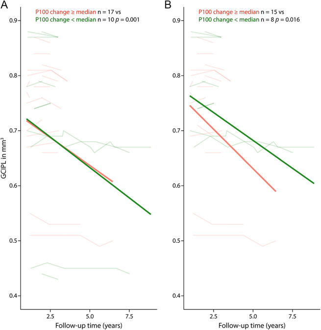
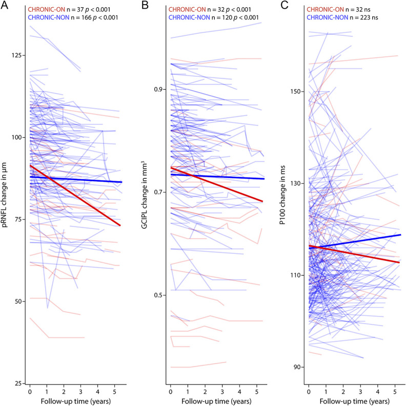
P100 latency change over the first year predicted subsequent GCIPL loss (36 months) across the entire chronic cohort ( = 0.001) and in (and driven by) the CHRONIC-NON subset ( = 0.019) but not in the CHRONIC-ON subset ( = 0.680). P100 latency and pRNFL were correlated at baseline (CHRONIC-NON = 0.004, CHRONIC-ON < 0.001), but change in P100 latency and pRNFL were not correlated. P100 latency did not differ longitudinally between protocols or centers.
VEP in non-ON eyes seems to be a promising marker of demyelination in RRMS and of potential prognostic value for subsequent retinal ganglion cell loss. This study also provides evidence that VEP may be a useful and reliable biomarker for multicenter studies.
随着视觉诱发电位(VEPs)作为临床试验中髓鞘的定量结果参数的应用日益增加,深入了解纵向 VEP 潜伏期变化及其对随后神经元丢失的预后潜力将是必要的。在这项纵向多中心研究中,我们评估了 VEP 潜伏期与复发缓解型多发性硬化症(RRMS)中光学相干断层扫描(OCT)测量的视网膜神经退行性变的相关性及其预后潜力。
我们纳入了 147 例 RRMS 患者的 293 只眼(年龄[岁,中位数±标准差]36±10,男性占 35%,随访[年,中位数{IQR}2.1{1.5-3.9}]):41 只眼基线时有≥6 个月的视神经炎(ON)病史(慢性 ON),252 只眼无 ON 病史(慢性 NON)。定量测量 P100 潜伏期(VEP)、黄斑联合神经节细胞和内丛状层体积(GCIPL)和视盘周围视网膜神经纤维层厚度(pRNFL)(OCT)。
第一年 P100 潜伏期的变化预测了整个慢性队列( = 0.001)和(并由)慢性 NON 亚组( = 0.019)在 36 个月时的后续 GCIPL 丢失,但在慢性 ON 亚组中则不然( = 0.680)。在慢性 NON 亚组( = 0.004,慢性 ON 组<0.001)和基线时,P100 潜伏期和 pRNFL 相关,但 P100 潜伏期和 pRNFL 的变化不相关。VEP 在不同协议或中心之间没有纵向差异。
非 ON 眼中的 VEP 似乎是 RRMS 脱髓鞘的有前途的标志物,对随后的视网膜神经节细胞丢失具有潜在的预后价值。本研究还提供了证据表明,VEP 可能是多中心研究的有用且可靠的生物标志物。