Coimbra Chemistry Center, Institute of Molecular Sciences (CQC-IMS), University of Coimbra, 3004-535 Coimbra, Portugal.
Department of Chemistry, Faculty of Sciences and Technology, University of Coimbra, 3004-535 Coimbra, Portugal.
Molecules. 2023 Feb 28;28(5):2241. doi: 10.3390/molecules28052241.
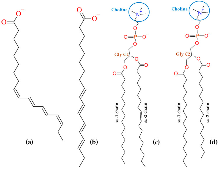
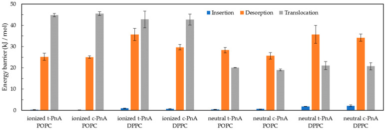
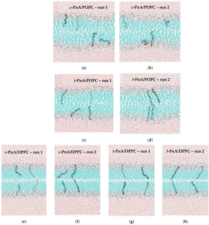
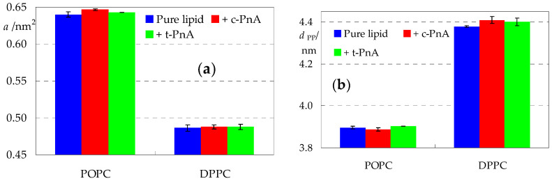
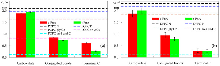
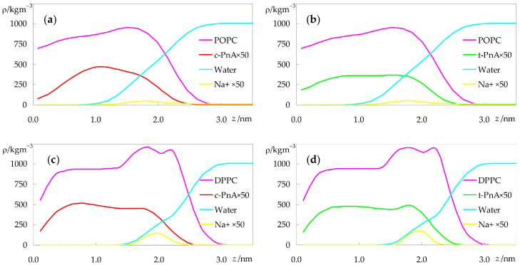
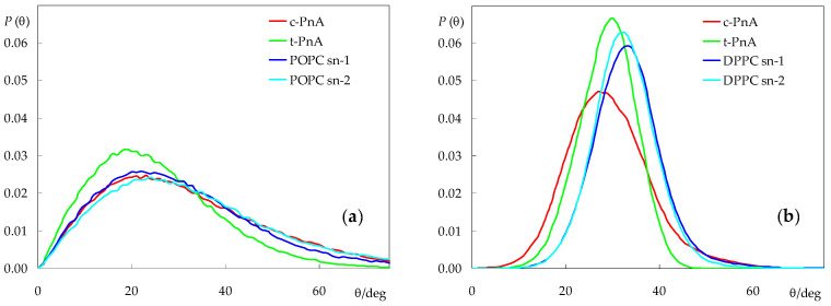
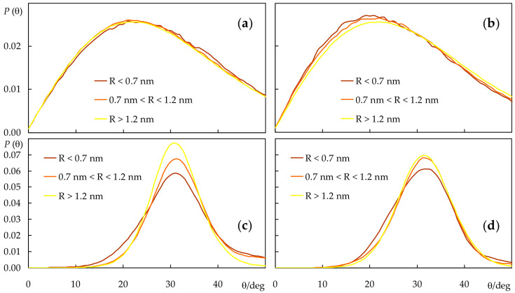
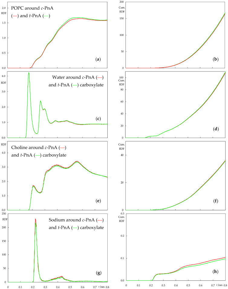
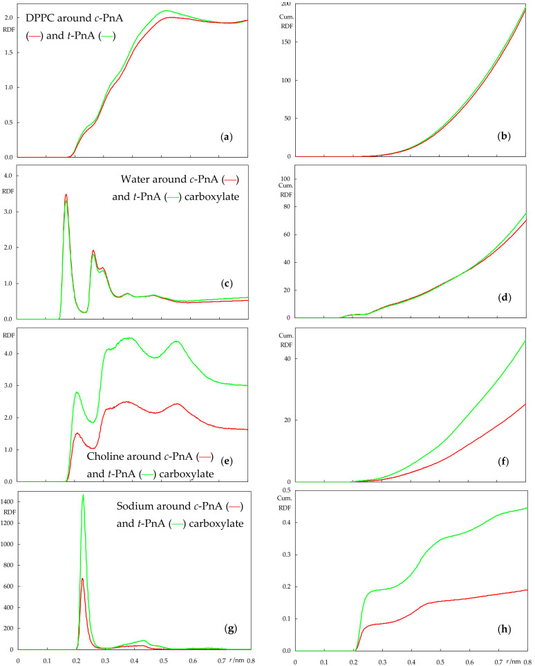
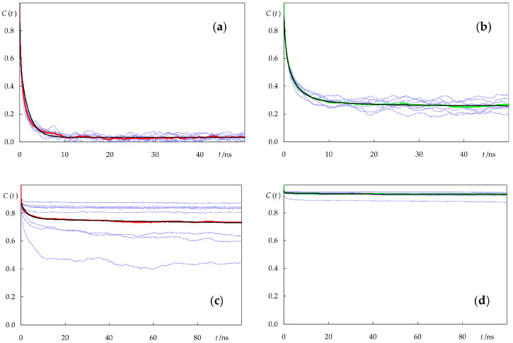
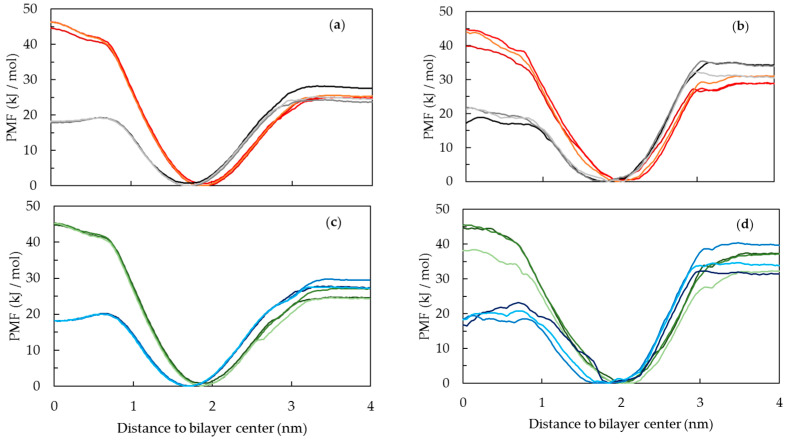
Fluorescence probes are indispensable tools in biochemical and biophysical membrane studies. Most of them possess extrinsic fluorophores, which often constitute a source of uncertainty and potential perturbation to the host system. In this regard, the few available intrinsically fluorescent membrane probes acquire increased importance. Among them, - and -parinaric acids (-PnA and -PnA, respectively) stand out as probes of membrane order and dynamics. These two compounds are long-chained fatty acids, differing solely in the configurations of two double bonds of their conjugated tetraene fluorophore. In this work, we employed all-atom and coarse-grained molecular dynamics simulations to study the behavior of -PnA and -PnA in lipid bilayers of 1-palmitoyl-2-oleoyl--glycero-3-phosphocholine (POPC) and 1,2-dipalmitoyl--glycero-3-phosphocholine (DPPC), representative of the liquid disordered and solid ordered lipid phases, respectively. All-atom simulations indicate that the two probes show similar location and orientation in the simulated systems, with the carboxylate facing the water/lipid interface and the tail spanning the membrane leaflet. The two probes establish interactions with the solvent and lipids to a similar degree in POPC. However, the almost linear -PnA molecules have tighter lipid packing around them, especially in DPPC, where they also interact more with positively charged lipid choline groups. Probably for these reasons, while both probes show similar partition (assessed from computed free energy profiles across bilayers) to POPC, -PnA clearly partitions more extensively than -PnA to the gel phase. -PnA also displays more hindered fluorophore rotation, especially in DPPC. Our results agree very well with experimental fluorescence data from the literature and allow deeper understanding of the behavior of these two reporters of membrane organization.
荧光探针是生物化学和生物物理膜研究中不可或缺的工具。它们大多具有外在的荧光团,这往往是宿主系统不确定性和潜在干扰的来源。在这方面,少数可用的内在荧光膜探针获得了更高的重要性。其中,- 和 - 帕纳酸(-PnA 和 -PnA,分别)作为膜有序性和动力学的探针脱颖而出。这两种化合物是长链脂肪酸,仅在其共轭四烯荧光团的两个双键的构型上有所不同。在这项工作中,我们使用全原子和粗粒分子动力学模拟来研究 -PnA 和 -PnA 在 1-棕榈酰基-2-油酰基--甘油-3-磷酸胆碱(POPC)和 1,2-二棕榈酰基--甘油-3-磷酸胆碱(DPPC)的脂质双层中的行为,分别代表液体无序相和固体有序相的脂质。全原子模拟表明,两种探针在模拟系统中表现出相似的位置和取向,羧酸根朝向水/脂质界面,尾部跨越膜叶。两种探针在 POPC 中与溶剂和脂质建立相似程度的相互作用。然而,几乎线性的 -PnA 分子周围的脂质包装更紧密,尤其是在 DPPC 中,它们与带正电荷的脂质胆碱基团也有更多的相互作用。可能由于这些原因,虽然两种探针都显示出与 POPC 相似的分配(从跨双层计算的自由能曲线评估),但 -PnA 明显比 -PnA 更广泛地分配到凝胶相。-PnA 的荧光团旋转也受到更多的阻碍,尤其是在 DPPC 中。我们的结果与文献中的实验荧光数据非常吻合,使我们能够更深入地了解这两种膜组织报告器的行为。