• 文献检索
  • 文档翻译
  • 深度研究
  • 学术资讯
  • Suppr Zotero 插件Zotero 插件
  • 邀请有礼
  • 套餐&价格
  • 历史记录
应用&插件
Suppr Zotero 插件Zotero 插件浏览器插件Mac 客户端Windows 客户端微信小程序
定价
高级版会员购买积分包购买API积分包
服务
文献检索文档翻译深度研究API 文档MCP 服务
关于我们
关于 Suppr公司介绍联系我们用户协议隐私条款
关注我们

Suppr 超能文献

核心技术专利:CN118964589B侵权必究
粤ICP备2023148730 号-1Suppr @ 2026

文献检索

告别复杂PubMed语法,用中文像聊天一样搜索,搜遍4000万医学文献。AI智能推荐,让科研检索更轻松。

立即免费搜索

文件翻译

保留排版,准确专业,支持PDF/Word/PPT等文件格式,支持 12+语言互译。

免费翻译文档

深度研究

AI帮你快速写综述,25分钟生成高质量综述,智能提取关键信息,辅助科研写作。

立即免费体验

乳腺癌监测中的对比增强乳腺摄影。

Contrast enhanced mammography in breast cancer surveillance.

机构信息

The Royal Melbourne Hospital, Grattan Street, Parkville, Melbourne, 3101, Australia.

Daffodil Centre, The University of Sydney, a joint venture with Cancer Council New South Wales, Sydney, Australia.

出版信息

Breast Cancer Res Treat. 2023 Jun;199(2):221-230. doi: 10.1007/s10549-023-06916-0. Epub 2023 Mar 25.

DOI:10.1007/s10549-023-06916-0
PMID:36966271
原文链接:https://pmc.ncbi.nlm.nih.gov/articles/PMC10175447/
Abstract

PURPOSE

Mammography (MG) is the standard imaging in surveillance of women with a personal history of breast cancer or DCIS (PHBC), supplemented with ultrasound. Contrast Enhanced Mammography (CEM) has higher sensitivity than MG and US. We report the performance of CEM compared with MG ± US.

METHODS

A retrospective study of patients undergoing their first surveillance CEM in an Australian hospital setting between June 2006 and October 2020. Cases where a patient was recalled for assessment were identified, recording radiology, pathology and treatment details. Blinded re-reading of recalled cases was performed to determine the contribution of contrast. Use of surveillance US across the board was assessed for the period.

RESULTS

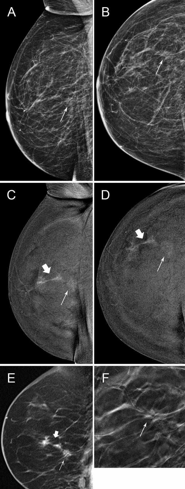
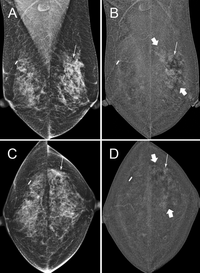
73/1191 (6.1%) patients were recalled. 35 (48%) were true positives (TP), with 26 invasive cancers and 9 cases of DCIS, while 38 (52%) were false positive (FP) with a positive predictive value (PPV) 47.9%. 32/73 were recalled due to MG findings, while 41/73 were only recalled due to Contrast. 14/73 had 'minimal signs' with a lesion identifiable on MG with knowledge of the contrast finding, while 27/73 were visible only with contrast. 41% (17/41) recalled due to contrast were TP. Contrast-only TPs were found with low and high mammographic density (MD). Screening breast US reduced by 55% in the year after CEM was implemented.

CONCLUSION

Compared to MG, CEM as a single surveillance modality for those with PHBC has higher sensitivity and comparable specificity, identifying additional malignant lesions that are clinically significant. Investigation of interval cancer and subsequent round outcomes is warranted.

摘要

目的

乳腺 X 线摄影术(MG)是具有乳腺癌个人史或 DCIS(PHBC)的女性监测的标准影像学方法,辅以超声检查。对比增强乳腺摄影术(CEM)比 MG 和 US 具有更高的敏感性。我们报告了 CEM 与 MG±US 的性能比较。

方法

回顾性研究了 2006 年 6 月至 2020 年 10 月期间在澳大利亚医院接受首次监测 CEM 的患者。确定了召回评估的患者病例,记录了放射学、病理学和治疗细节。对召回病例进行了盲法重读,以确定对比的贡献。评估了整个监测期间使用超声检查的情况。

结果

1191 例患者中有 73 例(6.1%)被召回。35 例(48%)为真阳性(TP),其中 26 例为浸润性癌,9 例为 DCIS,而 38 例(52%)为假阳性(FP),阳性预测值(PPV)为 47.9%。73 例中有 32 例因 MG 发现而被召回,而 41 例仅因对比而被召回。73 例中有 14 例(14/73)“有轻微迹象”,MG 上可识别病变,而对比检查发现有 27 例(27/73)仅通过对比检查发现。41%(17/41)因对比而召回的是 TP。在 CEM 实施后的一年中,筛查性乳腺超声检查减少了 55%。

结论

与 MG 相比,对于 PHBC 患者,CEM 作为单一监测方法具有更高的敏感性和相当的特异性,可识别出更多具有临床意义的恶性病变。需要对间期癌和后续轮次的结果进行研究。

https://cdn.ncbi.nlm.nih.gov/pmc/blobs/a48f/10175447/dd394fdfac52/10549_2023_6916_Fig3_HTML.jpg
https://cdn.ncbi.nlm.nih.gov/pmc/blobs/a48f/10175447/e095d0f7bee7/10549_2023_6916_Fig1_HTML.jpg
https://cdn.ncbi.nlm.nih.gov/pmc/blobs/a48f/10175447/85ad4038f1e5/10549_2023_6916_Fig2_HTML.jpg
https://cdn.ncbi.nlm.nih.gov/pmc/blobs/a48f/10175447/dd394fdfac52/10549_2023_6916_Fig3_HTML.jpg
https://cdn.ncbi.nlm.nih.gov/pmc/blobs/a48f/10175447/e095d0f7bee7/10549_2023_6916_Fig1_HTML.jpg
https://cdn.ncbi.nlm.nih.gov/pmc/blobs/a48f/10175447/85ad4038f1e5/10549_2023_6916_Fig2_HTML.jpg
https://cdn.ncbi.nlm.nih.gov/pmc/blobs/a48f/10175447/dd394fdfac52/10549_2023_6916_Fig3_HTML.jpg

相似文献

1
Contrast enhanced mammography in breast cancer surveillance.乳腺癌监测中的对比增强乳腺摄影。
Breast Cancer Res Treat. 2023 Jun;199(2):221-230. doi: 10.1007/s10549-023-06916-0. Epub 2023 Mar 25.
2
Contrast-enhanced mammography for surveillance in women with a personal history of breast cancer.对比增强乳腺摄影用于有乳腺癌个人史女性的监测。
Breast Cancer Res Treat. 2024 Nov;208(2):293-305. doi: 10.1007/s10549-024-07419-2. Epub 2024 Jul 4.
3
Contrast-Enhanced Mammography in Local Staging of Screen-Detected Breast Cancer.对比增强乳腺摄影在筛查性乳腺癌局部分期中的应用。
Ann Surg Oncol. 2024 Oct;31(10):6820-6830. doi: 10.1245/s10434-024-15848-y. Epub 2024 Jul 24.
4
Performance of artificial intelligence in 7533 consecutive prevalent screening mammograms from the BreastScreen Australia program.澳大利亚乳腺筛查计划中 7533 例连续现患筛查乳腺 X 线摄影的人工智能性能。
Eur Radiol. 2024 Jun;34(6):3947-3957. doi: 10.1007/s00330-023-10396-7. Epub 2023 Nov 13.
5
Contrast-enhanced mammography for the assessment of screening recalls: a two-centre study.对比增强乳腺摄影在筛查召回评估中的应用:一项两中心研究。
Eur Radiol. 2022 Nov;32(11):7388-7399. doi: 10.1007/s00330-022-08868-3. Epub 2022 Jun 1.
6
Role of contrast-enhanced mammography in the preoperative detection of ductal carcinoma in situ of the breasts: a comparison with low-energy image and magnetic resonance imaging.增强型乳腺钼靶摄影在乳腺导管原位癌术前检测中的作用:与低能影像和磁共振成像的比较。
Eur Radiol. 2024 May;34(5):3342-3351. doi: 10.1007/s00330-023-10312-z. Epub 2023 Oct 19.
7
Low-Dose, Contrast-Enhanced Mammography Compared to Contrast-Enhanced Breast MRI: A Feasibility Study.低剂量对比增强乳腺 X 光摄影与对比增强乳腺 MRI 对比:一项可行性研究。
J Magn Reson Imaging. 2020 Aug;52(2):589-595. doi: 10.1002/jmri.27079. Epub 2020 Feb 14.
8
Comparison of Abbreviated Breast MRI vs Digital Breast Tomosynthesis for Breast Cancer Detection Among Women With Dense Breasts Undergoing Screening.在接受筛查的致密型乳腺女性中,比较用于乳腺癌检测的简化乳腺 MRI 与数字乳腺断层合成术。
JAMA. 2020 Feb 25;323(8):746-756. doi: 10.1001/jama.2020.0572.
9
The PROCEM study protocol: Added value of preoperative contrast-enhanced mammography in staging of malignant breast lesions - a prospective randomized multicenter study.PROCEM 研究方案:术前对比增强乳腺摄影在恶性乳腺病变分期中的附加价值 - 一项前瞻性随机多中心研究。
BMC Cancer. 2021 Oct 18;21(1):1115. doi: 10.1186/s12885-021-08832-2.
10
Utility of Targeted Ultrasound to Predict Malignancy Among Lesions Detected on Contrast-Enhanced Digital Mammography.超声造影增强数字钼靶检查对良恶性病变的预测价值
AJR Am J Roentgenol. 2021 Sep;217(3):595-604. doi: 10.2214/AJR.20.24368. Epub 2020 Oct 7.

引用本文的文献

1
ESR Essentials: post-treatment breast cancer surveillance from mammography to a more personalised approach-practice recommendations by the European Society of Breast Imaging.红细胞沉降率要点:乳腺癌治疗后监测——从乳腺摄影到更个性化的方法——欧洲乳腺影像学会的实践建议
Eur Radiol. 2025 Aug 14. doi: 10.1007/s00330-025-11819-3.
2
ASBrS Keynote Address: Reimagining Stage 1 Breast Cancer-Evolution of Breast Cancer Management over the Last 120 Years.美国乳腺外科医师学会主题演讲:重塑早期乳腺癌——过去120年乳腺癌管理的演变
Ann Surg Oncol. 2025 Aug 7. doi: 10.1245/s10434-025-17849-x.
3
Preliminary Evaluation of Radiomics in Contrast-Enhanced Mammography for Prognostic Prediction of Breast Cancer.

本文引用的文献

1
Evaluation of Adjunctive Ultrasonography for Breast Cancer Detection Among Women Aged 40-49 Years With Varying Breast Density Undergoing Screening Mammography: A Secondary Analysis of a Randomized Clinical Trial.乳腺钼靶筛查中不同乳腺密度的40-49岁女性乳腺癌检测辅助超声检查的评估:一项随机临床试验的二次分析
JAMA Netw Open. 2021 Aug 2;4(8):e2121505. doi: 10.1001/jamanetworkopen.2021.21505.
2
Interval Cancers after Negative Supplemental Screening Breast MRI Results in Women with a Personal History of Breast Cancer.乳腺癌病史女性阴性补充乳腺 MRI 筛查结果后的间期癌。
Radiology. 2021 Aug;300(2):314-323. doi: 10.1148/radiol.2021203074. Epub 2021 Jun 8.
3
对比增强乳腺钼靶摄影中影像组学对乳腺癌预后预测的初步评估
Cancers (Basel). 2025 Jun 10;17(12):1926. doi: 10.3390/cancers17121926.
4
Contrast-enhanced mammography for breast cancer detection and diagnosis with high concentration iodinated contrast medium.使用高浓度碘化造影剂的对比增强乳腺摄影术用于乳腺癌的检测和诊断。
Insights Imaging. 2025 Jun 14;16(1):124. doi: 10.1186/s13244-025-01994-8.
5
Imaging in Interventional Radiology: Breast Cryoablation.介入放射学中的影像学:乳腺冷冻消融术
Semin Intervent Radiol. 2025 Mar 27;42(1):101-113. doi: 10.1055/s-0045-1806738. eCollection 2025 Feb.
6
Evaluation of contrast-enhanced mammography (CEM) in the preoperative staging of breast cancer: large-scale single center experience, update to 1005 cases.对比增强乳腺钼靶摄影(CEM)在乳腺癌术前分期中的评估:大规模单中心经验,病例更新至1005例
Radiol Med. 2025 Mar 28. doi: 10.1007/s11547-025-02009-8.
7
Breast MRI to Screen Women With Extremely Dense Breasts.乳腺磁共振成像用于筛查乳腺极度致密的女性。
J Magn Reson Imaging. 2025 Jul;62(1):58-72. doi: 10.1002/jmri.29716. Epub 2025 Jan 24.
8
Contrast-enhanced mammography improves patient access to functional breast imaging.对比增强乳腺钼靶摄影改善了患者获得功能性乳腺成像的机会。
J Med Imaging Radiat Oncol. 2025 Feb;69(1):46-61. doi: 10.1111/1754-9485.13789. Epub 2024 Oct 31.
9
Contrast-enhanced mammography for surveillance in women with a personal history of breast cancer.对比增强乳腺摄影用于有乳腺癌个人史女性的监测。
Breast Cancer Res Treat. 2024 Nov;208(2):293-305. doi: 10.1007/s10549-024-07419-2. Epub 2024 Jul 4.
10
Addition of Contrast-enhanced Mammography to Tomosynthesis for Breast Cancer Detection in Women with a Personal History of Breast Cancer: Prospective TOCEM Trial Interim Analysis.增强型乳腺 X 光摄影术联合断层合成乳腺摄影术在有乳腺癌个人病史的女性乳腺癌检测中的应用:前瞻性 TOCEM 试验中期分析。
Radiology. 2024 Apr;311(1):e231991. doi: 10.1148/radiol.231991.
Digital Mammography and Breast Tomosynthesis Performance in Women with a Personal History of Breast Cancer, 2007-2016.
2007-2016 年有乳腺癌个人病史女性的数字乳腺 X 线摄影与乳腺断层合成性能
Radiology. 2021 Aug;300(2):290-300. doi: 10.1148/radiol.2021204581. Epub 2021 May 18.
4
Contrast-Enhanced Mammography for Screening Women after Breast Conserving Surgery.保乳术后女性筛查的对比增强乳腺造影术。
Cancers (Basel). 2020 Nov 24;12(12):3495. doi: 10.3390/cancers12123495.
5
Breast ultrasound in breast cancer surveillance; incremental cancers found at what cost?乳腺癌监测中的乳腺超声:以什么为代价发现的新增癌症?
Breast. 2020 Dec;54:272-277. doi: 10.1016/j.breast.2020.11.007. Epub 2020 Nov 11.
6
Supplemental breast cancer-screening ultrasonography in women with dense breasts: a systematic review and meta-analysis.致密型乳腺女性补充性乳腺癌筛查超声检查:系统评价和荟萃分析。
Br J Cancer. 2020 Aug;123(4):673-688. doi: 10.1038/s41416-020-0928-1. Epub 2020 Jun 12.
7
A meta-analysis of contrast-enhanced spectral mammography versus MRI in the diagnosis of breast cancer.对比增强光谱乳腺摄影与 MRI 在乳腺癌诊断中的荟萃分析。
Thorac Cancer. 2020 Jun;11(6):1423-1432. doi: 10.1111/1759-7714.13400. Epub 2020 Mar 31.
8
Supplemental Breast US Screening in Women with a Personal History of Breast Cancer: A Matched Cohort Study.乳腺癌病史女性的补充性乳腺超声筛查:一项匹配队列研究。
Radiology. 2020 Apr;295(1):54-63. doi: 10.1148/radiol.2020191691. Epub 2020 Feb 25.
9
Supplemental MRI Screening for Women with Extremely Dense Breast Tissue.致密型乳腺女性的补充性磁共振成像筛查。
N Engl J Med. 2019 Nov 28;381(22):2091-2102. doi: 10.1056/NEJMoa1903986.
10
Mammographic Density and Screening Sensitivity, Breast Cancer Incidence and Associated Risk Factors in Danish Breast Cancer Screening.丹麦乳腺癌筛查中的乳腺X线密度与筛查敏感性、乳腺癌发病率及相关危险因素
J Clin Med. 2019 Nov 19;8(11):2021. doi: 10.3390/jcm8112021.