Coroamă Diana Mihaela, Dioșan Laura, Telecan Teodora, Andras Iulia, Crișan Nicolae, Medan Paul, Andreica Anca, Caraiani Cosmin, Lebovici Andrei, Boca Bianca, Bálint Zoltán
Faculty of Mathematics and Computer Science, Babeș-Bolyai University, Cluj-Napoca, Romania.
Department of Urology, Faculty of Medicine, Iuliu Hațieganu University of Medicine and Pharmacy, Cluj-Napoca, Romania.
Front Oncol. 2023 Mar 9;13:1096136. doi: 10.3389/fonc.2023.1096136. eCollection 2023.
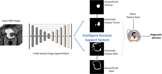
Bladder magnetic resonance imaging (MRI) has been recently integrated in the diagnosis pathway of bladder cancer. However, automatic recognition of suspicious lesions is still challenging. Thus, development of a solution for proper delimitation of the tumor and its separation from the healthy tissue is of primordial importance. As a solution to this unmet medical need, we aimed to develop an artificial intelligence-based decision support system, which automatically segments the bladder wall and the tumor as well as any suspect area from the 3D MRI images.
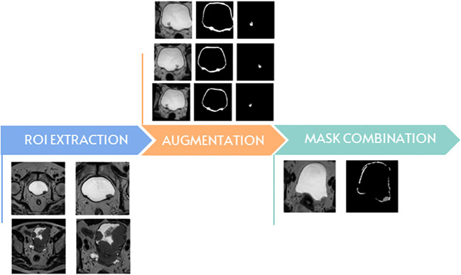
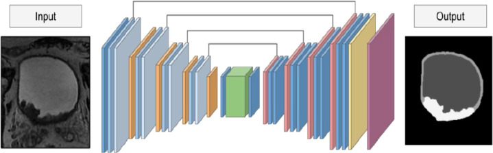
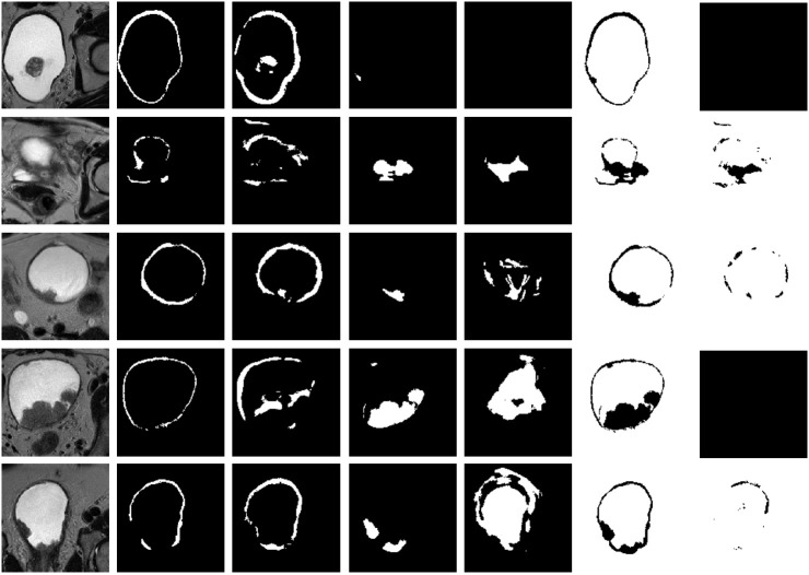
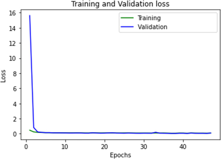
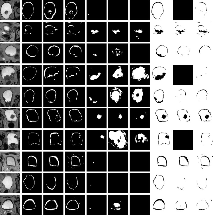
We retrospectively assessed all patients diagnosed with bladder cancer, who underwent MRI at our department (n=33). All examinations were performed using a 1.5 Tesla MRI scanner. All images were reviewed by two radiologists, who performed manual segmentation of the bladder wall and all lesions. First, the performance of our fully automated end-to-end segmentation model based on a 3D U-Net architecture (by considering various depths of 4, 5 or 6 blocks) trained in two data augmentation scenarios (on 5 and 10 augmentation datasets per original data, respectively) was tested. Second, two learning setups were analyzed by training the segmentation algorithm with 7 and 14 MRI original volumes, respectively.
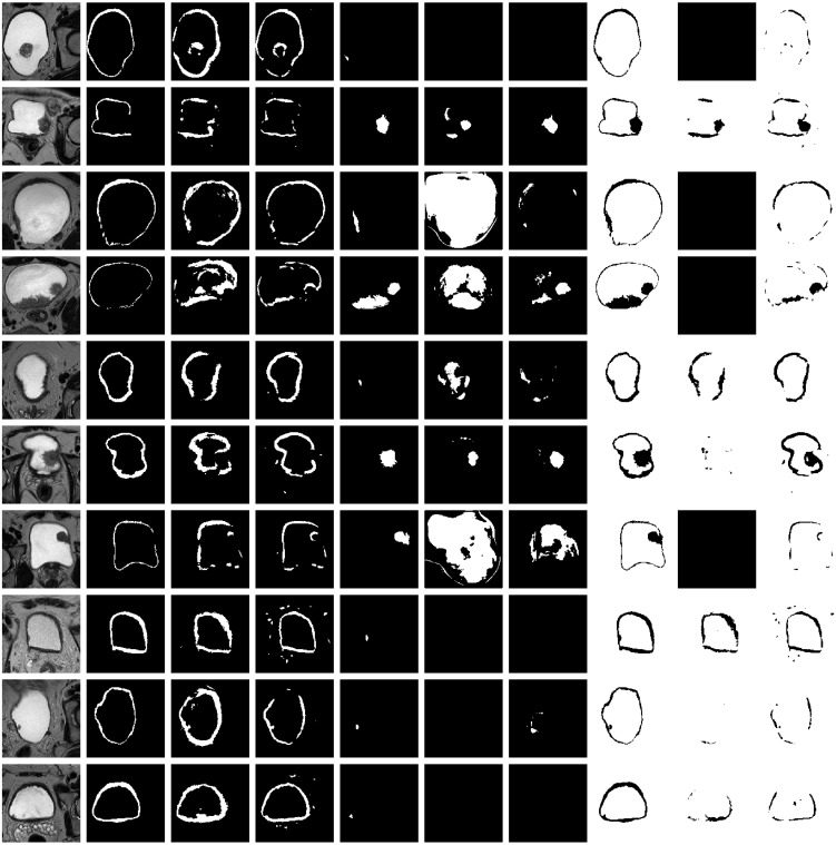
We obtained a Dice-based performance over 0.878 for automatic segmentation of bladder wall and tumors, as compared to manual segmentation. A larger training dataset using 10 augmentations for 7 patients could further improve the results of the U-Net-5 model (0.902 Dice coefficient at image level). This model performed best in terms of automated segmentation of bladder, as compared to U-Net-4 and U-Net-6. However, in this case increased time for learning was needed as compared to U-Net-4. We observed that an extended dataset for training led to significantly improved segmentation of the bladder wall, but not of the tumor.
We developed an intelligent system for bladder tumors automated diagnostic, that uses a deep learning model to segment both the bladder wall and the tumor. As a conclusion, low complexity networks, with less than five-layers U-Net architecture are feasible and show good performance for automatic 3D MRI image segmentation in patients with bladder tumors.
膀胱磁共振成像(MRI)最近已被纳入膀胱癌的诊断流程。然而,自动识别可疑病变仍然具有挑战性。因此,开发一种用于正确界定肿瘤并将其与健康组织分离的解决方案至关重要。作为满足这一未满足的医疗需求的解决方案,我们旨在开发一种基于人工智能的决策支持系统,该系统可从3D MRI图像中自动分割膀胱壁、肿瘤以及任何可疑区域。
我们回顾性评估了所有在我院接受MRI检查并被诊断为膀胱癌的患者(n = 33)。所有检查均使用1.5特斯拉MRI扫描仪进行。所有图像均由两位放射科医生进行评估,他们对膀胱壁和所有病变进行了手动分割。首先,测试了我们基于3D U-Net架构的全自动端到端分割模型(考虑4、5或6个块的各种深度)在两种数据增强场景(分别在每个原始数据的5个和10个增强数据集上)下的性能。其次,通过分别用7个和14个MRI原始体积训练分割算法,分析了两种学习设置。
与手动分割相比,我们在膀胱壁和肿瘤的自动分割方面获得了基于Dice的性能超过0.878。对7名患者使用10次增强的更大训练数据集可以进一步改善U-Net-5模型的结果(图像水平的Dice系数为0.902)。与U-Net-4和U-Net-6相比,该模型在膀胱自动分割方面表现最佳。然而,在这种情况下,与U-Net-4相比需要更长的学习时间。我们观察到,扩展的训练数据集导致膀胱壁的分割有显著改善,但肿瘤的分割没有改善。
我们开发了一种用于膀胱肿瘤自动诊断的智能系统,该系统使用深度学习模型分割膀胱壁和肿瘤。总之,具有少于五层U-Net架构的低复杂度网络是可行的,并且在膀胱肿瘤患者的自动3D MRI图像分割中表现出良好的性能。