Department of Gastroenterological Surgery, Nagoya City University Graduate School of Medical Sciences, 1 Kawasumi, Mizuho-cho, Mizuho-ku, Nagoya, Aichi, 4678601, Japan.
J Med Case Rep. 2023 Mar 28;17(1):127. doi: 10.1186/s13256-023-03828-x.
Neuroendocrine tumors of the minor papilla are very rare, and only 20 cases have been reported in the literature. Neuroendocrine carcinoma of the minor papilla with pancreas divisum has not been reported previously, making this the first reported case. Neuroendocrine tumors of the minor papilla have been reported in association with pancreas divisum in about 50% of cases reported in the literature. We herein present our case of neuroendocrine carcinoma of the minor papilla with pancreas divisum in a 75-year-old male with a systematic literature review of the previous 20 reports of neuroendocrine tumors of the minor papilla.
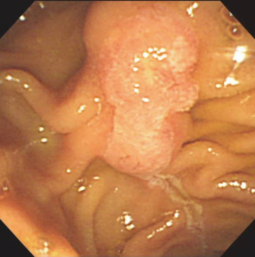
A 75-year-old Asian man was referred to our hospital for evaluation of dilation of the main pancreatic duct noted on abdominal ultrasonography. Magnetic resonance cholangiopancreatography and endoscopic retrograde cholangiopancreatography showed a dilated dorsal pancreatic duct, which was not connected to the ventral pancreatic duct; however, it opened to the minor papilla, indicating pancreas divisum. The common bile duct had no communication with the pancreatic main duct and opened to the ampulla of Vater. A contrast-enhanced computed tomography scan showed a 12-mm hypervascular mass near the ampulla of Vater. Endoscopic ultrasonography showed a defined hypoechoic mass in the minor papilla with no invasion. The biopsies performed at the previous hospital found adenocarcinoma. The patient underwent a subtotal stomach-preserving pancreaticoduodenectomy. The pathological diagnosis was neuroendocrine carcinoma. At the 15-year follow-up visit, the patient was doing well with no evidence of tumor recurrence.
In our case, because the tumor was discovered during a medical check-up relatively early in the course of disease, the patient was doing well at the 15-year follow-up visit, with no evidence of tumor recurrence. Diagnosing a tumor of the minor papilla is very difficult because of the relatively small size and submucosal location. Carcinoids and endocrine cell micronests in the minor papilla occur more frequently than generally thought. It is very important to include neuroendocrine tumors of the minor papilla in the differential diagnosis of patients with recurrent pancreatitis or pancreatitis of unknown cause, especially for patients with pancreas divisum.
小乳头神经内分泌肿瘤非常罕见,文献中仅报道了 20 例。此前从未报道过小乳头神经内分泌癌伴胰腺分裂症,因此这是首例报道。文献报道的小乳头神经内分泌肿瘤中,约有 50%与胰腺分裂症有关。我们在此报告一例小乳头神经内分泌癌伴胰腺分裂症的病例,并对之前 20 例小乳头神经内分泌肿瘤病例进行了系统的文献回顾。
一名 75 岁的亚洲男性因腹部超声检查发现主胰管扩张而被转诊至我院。磁共振胰胆管成像和内镜逆行胰胆管造影显示背侧胰管扩张,与腹侧胰管不相连;然而,它通向小乳头,提示胰腺分裂症。胆总管与胰总管无沟通,开口于 Vater 壶腹。增强 CT 扫描显示 Vater 壶腹附近有一个 12mm 的富血管性肿块。内镜超声显示小乳头有一个界限清楚的低回声肿块,无侵犯。前一家医院的活检发现腺癌。患者接受了保留胃的胰十二指肠切除术。病理诊断为神经内分泌癌。在 15 年的随访中,患者情况良好,无肿瘤复发迹象。
在我们的病例中,由于肿瘤在疾病早期的体检中被发现,患者在 15 年的随访中情况良好,无肿瘤复发迹象。由于肿瘤体积较小且位于黏膜下,因此诊断小乳头肿瘤非常困难。小乳头的类癌和内分泌细胞微巢比普遍认为的更为常见。对于复发性胰腺炎或不明原因胰腺炎的患者,特别是胰腺分裂症患者,将小乳头神经内分泌肿瘤纳入鉴别诊断非常重要。