Wagner Matthias W, Rafful Patricia P, Vidarsson Logi, Ertl-Wagner Birgit B
Division of Neuroradiology, Department of Diagnostic Imaging, The Hospital for Sick Children (SickKids), Toronto, ON M5G 1X8, Canada.
Neurosciences & Mental Health Research Program, SickKids Research Institute, Toronto, ON M5G 1X8, Canada.
Children (Basel). 2023 Feb 27;10(3):477. doi: 10.3390/children10030477.
Literature is scarce regarding volumetric measures of limbic system components across the pediatric age range. The purpose of this study is to remedy this scarcity by reporting continuous volumetric measurements of limbic system components, and to provide consistent stratification data including age-related trajectories and sex-related differences in the pediatric age range in order to improve the recognition of structural variations that might reflect pathology.
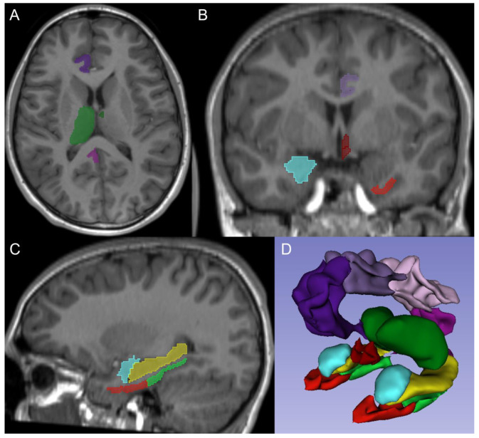
In this retrospective study, MRI sequences of children with normal clinical MRI examinations of the brain acquired between January 2010 and December 2019 were included. Isotropic 3D T1-weighted were processed using FreeSurfer version 7.3. Total brain volume and volumes of the limbic system including the hippocampus, parahippocampal gyrus, amygdala, hypothalamus, cingulate gyrus, entorhinal cortex, anteroventral thalamic nucleus, and whole thalamus were assessed. Parcellated output was displayed with the respective label map overlay and images were visually inspected for accuracy of regional segmentation results. Continuous data are provided as mean and standard deviation with quadratic trendlines and as mean and 95% confidence intervals. Categorical data are presented as integers and percentages (%).
A total of 724 children (401 female, 55.4%), with a mean age at time of MRI of 10.9 ± 4.2 years (range: 1.9-18.2 years), were included in the study. For females, the total brain volume increased from 955 ± 70 mL at the age of 2-3 years to 1140 ± 110 mL at the age of 17-18 years. Similarly, the total brain volume increased for males from 1004 ± 83 mL to 1263 ± 96 mL. The maximum volume was noted at 11-12 years for females (1188 ± 90 mL) and at 14-15 years for males (1310 ± 159 mL). Limbic system structures reached their peak volume more commonly between the 13-14 years to 17-18 years age groups. The male cingulate gyrus, entorhinal cortex, and anteroventral thalamic nucleus reached peak volume before or at 9-10 years.
This study provides unique age- and sex-specific volumes of the components of the limbic system throughout the pediatric age range to serve as normal values in comparative studies. Quantification of volumetric abnormalities of the limbic system on brain MRI may offer insights into phenotypical variations of diseases and may help elucidate new pathological phenotypes.
关于整个儿童年龄范围内边缘系统组成部分的体积测量的文献较少。本研究的目的是通过报告边缘系统组成部分的连续体积测量来弥补这一不足,并提供一致的分层数据,包括儿童年龄范围内与年龄相关的轨迹和与性别相关的差异,以提高对可能反映病理学的结构变异的识别。
在这项回顾性研究中,纳入了2010年1月至2019年12月期间接受脑部临床MRI检查正常的儿童的MRI序列。使用FreeSurfer 7.3版本处理各向同性3D T1加权图像。评估全脑体积以及边缘系统的体积,包括海马体、海马旁回、杏仁核、下丘脑、扣带回、内嗅皮质、丘脑前腹核和整个丘脑。分割后的输出结果与相应的标签图叠加显示,并对图像进行视觉检查以确保区域分割结果的准确性。连续数据以平均值和标准差以及二次趋势线的形式提供,同时也以平均值和95%置信区间的形式提供。分类数据以整数和百分比(%)表示。
本研究共纳入724名儿童(401名女性,占55.4%),MRI检查时的平均年龄为10.9±4.2岁(范围:1.9 - 18.2岁)。对于女性,全脑体积从2 - 3岁时的955±70 mL增加到17 - 18岁时的1140±110 mL。同样,男性的全脑体积从1004±83 mL增加到1263±96 mL。女性在11 - 12岁时达到最大体积(1188±90 mL),男性在14 - 15岁时达到最大体积(1310±159 mL)。边缘系统结构在13 - 14岁至17 - 18岁年龄组之间更常达到其峰值体积。男性的扣带回、内嗅皮质和丘脑前腹核在9 - 10岁之前或9 - 10岁时达到峰值体积。
本研究提供了整个儿童年龄范围内边缘系统各组成部分独特的年龄和性别特异性体积,可作为比较研究中的正常值。通过脑MRI对边缘系统体积异常进行量化,可能有助于深入了解疾病的表型变异,并可能有助于阐明新的病理表型。