FinnBrain Birth Cohort Study, Turku Brain and Mind Center, Department of Clinical Medicine, University of Turku, Turku, Finland.
Department of Psychiatry, Turku University Hospital, University of Turku, Turku, Finland.
Eur J Neurosci. 2022 Sep;56(5):4619-4641. doi: 10.1111/ejn.15761. Epub 2022 Jul 18.
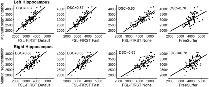
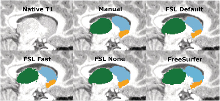
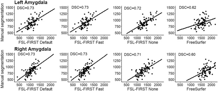
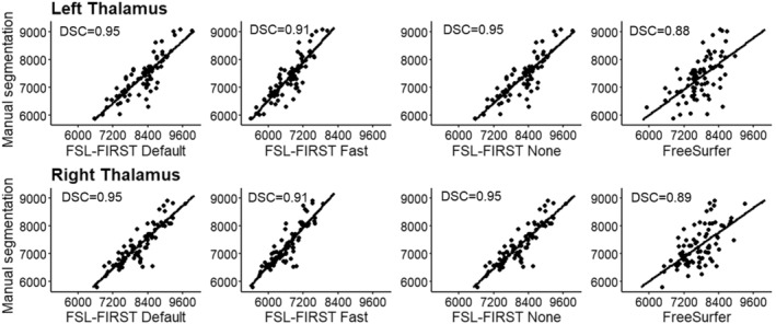
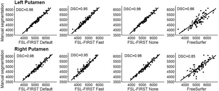
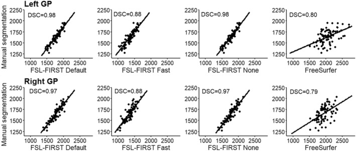
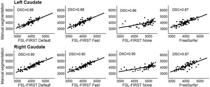
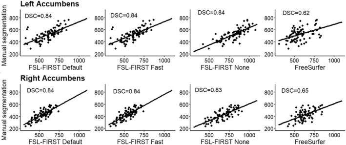
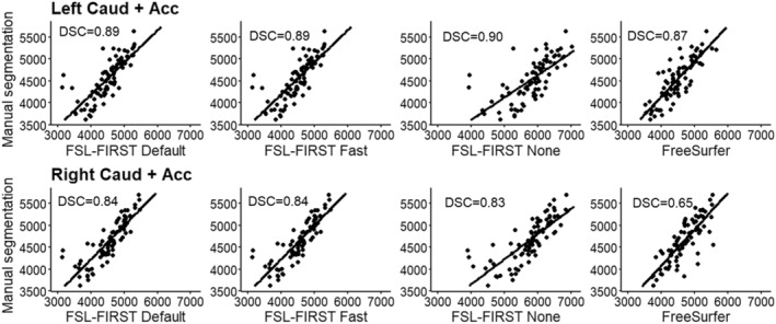
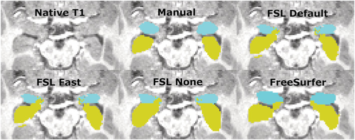
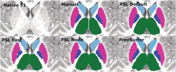
Developing accurate subcortical volumetric quantification tools is crucial for neurodevelopmental studies, as they could reduce the need for challenging and time-consuming manual segmentation. In this study, the accuracy of two automated segmentation tools, FSL-FIRST (with three different boundary correction settings) and FreeSurfer, were compared against manual segmentation of the hippocampus and subcortical nuclei, including the amygdala, thalamus, putamen, globus pallidus, caudate and nucleus accumbens, using volumetric and correlation analyses in 80 5-year-olds. Both FSL-FIRST and FreeSurfer overestimated the volume on all structures except the caudate, and the accuracy varied depending on the structure. Small structures such as the amygdala and nucleus accumbens, which are visually difficult to distinguish, produced significant overestimations and weaker correlations with all automated methods. Larger and more readily distinguishable structures such as the caudate and putamen produced notably lower overestimations and stronger correlations. Overall, the segmentations performed by FSL-FIRST's default pipeline were the most accurate, whereas FreeSurfer's results were weaker across the structures. In line with prior studies, the accuracy of automated segmentation tools was imperfect with respect to manually defined structures. However, apart from amygdala and nucleus accumbens, FSL-FIRST's agreement could be considered satisfactory (Pearson correlation > 0.74, intraclass correlation coefficient (ICC) > 0.68 and Dice score coefficient (DSC) > 0.87) with highest values for the striatal structures (putamen, globus pallidus, caudate) (Pearson correlation > 0.77, ICC > 0.87 and DSC > 0.88, respectively). Overall, automated segmentation tools do not always provide satisfactory results, and careful visual inspection of the automated segmentations is strongly advised.
开发准确的皮质下容积量化工具对于神经发育研究至关重要,因为它们可以减少对具有挑战性和耗时的手动分割的需求。在这项研究中,比较了两种自动分割工具(FSL-FIRST(具有三种不同的边界校正设置)和 FreeSurfer)与手动分割海马体和皮质下核(包括杏仁核、丘脑、壳核、苍白球、尾状核和伏隔核)的准确性,使用容积和相关性分析在 80 名 5 岁儿童中进行。FSL-FIRST 和 FreeSurfer 都高估了所有结构的体积,除了尾状核,其准确性取决于结构。视觉上难以区分的小结构,如杏仁核和伏隔核,产生了显著的高估和与所有自动方法较弱的相关性。较大且更容易区分的结构,如尾状核和壳核,产生的高估较小,相关性较强。总体而言,FSL-FIRST 默认管道执行的分割是最准确的,而 FreeSurfer 的结果在所有结构中都较弱。与先前的研究一致,自动分割工具的准确性对于手动定义的结构并不完美。然而,除了杏仁核和伏隔核之外,FSL-FIRST 的一致性可以被认为是令人满意的(Pearson 相关系数>0.74,组内相关系数(ICC)>0.68 和骰子分数系数(DSC)>0.87),最高值为纹状体结构(壳核、苍白球、尾状核)(Pearson 相关系数>0.77,ICC>0.87 和 DSC>0.88,分别)。总体而言,自动分割工具并不总是提供令人满意的结果,强烈建议仔细检查自动分割。