Liffner Benjamin, Cepeda Diaz Ana Karla, Blauwkamp James, Anaguano David, Frölich Sonja, Muralidharan Vasant, Wilson Danny W, Dvorin Jeffrey, Absalon Sabrina
Department of Pharmacology and Toxicology, Indiana University School of Medicine, Indianapolis, IN, USA.
Biological and Biomedical Sciences, Harvard Medical School, Boston MA, USA.
bioRxiv. 2023 Oct 9:2023.03.22.533773. doi: 10.1101/2023.03.22.533773.
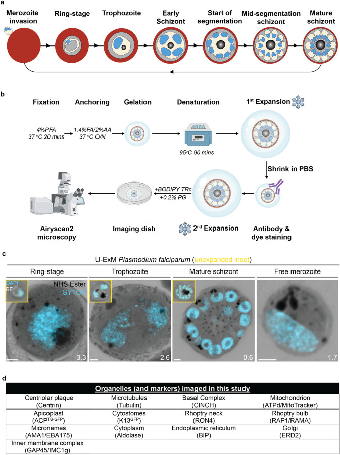
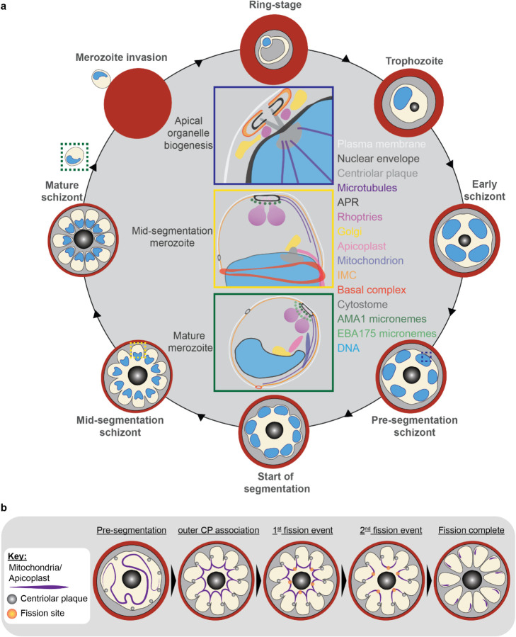
Apicomplexan parasites exhibit tremendous diversity in much of their fundamental cell biology, but study of these organisms using light microscopy is often hindered by their small size. Ultrastructural expansion microscopy (U-ExM) is a microscopy preparation method that physically expands the sample ~4.5x. Here, we apply U-ExM to the human malaria parasite during the asexual blood stage of its lifecycle to understand how this parasite is organized in three-dimensions. Using a combination of dye-conjugated reagents and immunostaining, we have catalogued 13 different structures or organelles across the intraerythrocytic development of this parasite and made multiple observations about fundamental parasite cell biology. We describe that the outer centriolar plaque and its associated proteins anchor the nucleus to the parasite plasma membrane during mitosis. Furthermore, the rhoptries, Golgi, basal complex, and inner membrane complex, which form around this anchoring site while nuclei are still dividing, are concurrently segregated and maintain an association to the outer centriolar plaque until the start of segmentation. We also show that the mitochondrion and apicoplast undergo sequential fission events while maintaining an association with the outer centriolar plaque during cytokinesis. Collectively, this study represents the most detailed ultrastructural analysis of during its intraerythrocytic development to date, and sheds light on multiple poorly understood aspects of its organelle biogenesis and fundamental cell biology.
顶复门寄生虫在其许多基本细胞生物学方面表现出巨大的多样性,但使用光学显微镜对这些生物体进行研究常常因其体积小而受到阻碍。超微结构扩张显微镜(U-ExM)是一种将样品物理性地扩大约4.5倍的显微镜制备方法。在此,我们将U-ExM应用于人类疟原虫生命周期中的无性血液阶段,以了解该寄生虫在三维空间中的组织方式。通过结合染料偶联试剂和免疫染色,我们梳理了该寄生虫在红细胞内发育过程中的13种不同结构或细胞器,并对疟原虫基本细胞生物学进行了多项观察。我们描述了在有丝分裂期间,外部中心粒斑块及其相关蛋白将细胞核锚定在寄生虫质膜上。此外,在细胞核仍在分裂时围绕该锚定位点形成的棒状体、高尔基体、基部复合体和内膜复合体,会同时被分离,并与外部中心粒斑块保持联系,直到开始分裂。我们还表明,线粒体和质体在胞质分裂期间经历连续的分裂事件,同时与外部中心粒斑块保持联系。总体而言,这项研究代表了迄今为止对疟原虫红细胞内发育最详细的超微结构分析,并揭示了其细胞器生物发生和基本细胞生物学中多个了解不足的方面。