Nakashima Takema, Morimoto Tadatsugu, Hashimoto Akira, Kii Sakumo, Tsukamoto Masatsugu, Miyamoto Hiroshi, Todo Mitsugu, Sonohata Motoki, Mawatari Masaaki
Department of Orthopaedic Surgery, Faculty of Medicine Saga University Saga Japan.
Department of Pathology and Microbiology, Faculty of Medicine Saga University Saga Japan.
JOR Spine. 2022 Nov 29;6(1):e1236. doi: 10.1002/jsp2.1236. eCollection 2023 Mar.
The use of spinal instrumentation is an established risk factor for postoperative infection. To address this problem, we prepared silver-containing hydroxyapatite coating, consisting of highly osteoconductive hydroxyapatite interfused with silver. The technology has been adopted for total hip arthroplasty. Silver-containing hydroxyapatite coating has been reported to have good biocompatibility and low toxicity. However, no studies about applying this coating in spinal surgery have addressed the osteoconductivity and direct neurotoxicity to the spinal cord of silver-containing hydroxyapatite cages in spinal interbody fusion.
In this study, we evaluated the osteoconductivity and neurotoxicity of silver-containing hydroxyapatite-coated implants in rats.
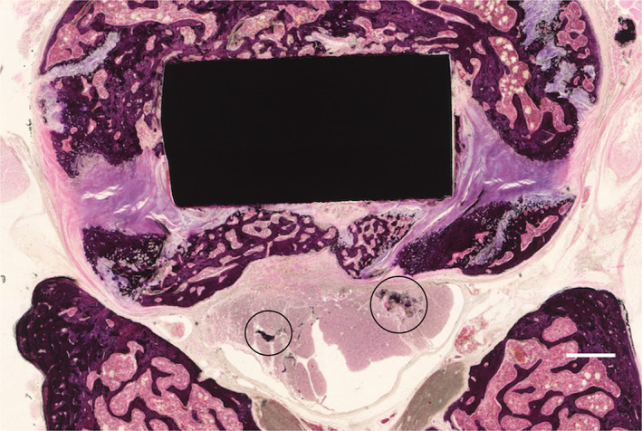
MATERIALS & METHODS: Titanium (non-coated, hydroxyapatite-coated, and silver-containing hydroxyapatite-coated) interbody cages were inserted into the spine for anterior lumbar fusion. At 8 weeks postoperatively, micro-computed tomography and histology were performed to evaluate the osteoconductivity of the cage. Inclined plane test and toe pinch test were performed postoperatively to assess neurotoxicity.
Micro-computed tomography data indicated no significant difference in bone volume/total volume among the three groups. Histologically, the hydroxyapatite-coated and silver-containing hydroxyapatite-coated groups showed significantly higher bone contact rate than that of the titanium group. In contrast, there was no significant difference in bone formation rate among the three groups. Data of inclined plane and toe pinch test showed no significant loss of motor and sensory function in the three groups. Furthermore, there was no degeneration, necrosis, or accumulation of silver in the spinal cord on histology.
This study suggests that silver-hydroxyapatite-coated interbody cages produce good osteoconductivity and are not associated with direct neurotoxicity.
脊柱内固定器械的使用是术后感染的一个既定风险因素。为解决这一问题,我们制备了含银羟基磷灰石涂层,它由与银融合的高骨传导性羟基磷灰石组成。该技术已应用于全髋关节置换术。据报道,含银羟基磷灰石涂层具有良好的生物相容性和低毒性。然而,关于在脊柱手术中应用这种涂层的研究尚未涉及含银羟基磷灰石椎间融合器在脊柱椎间融合中的骨传导性和对脊髓的直接神经毒性。
在本研究中,我们评估了含银羟基磷灰石涂层植入物在大鼠体内的骨传导性和神经毒性。
将钛(未涂层、羟基磷灰石涂层和含银羟基磷灰石涂层)椎间融合器植入脊柱进行前路腰椎融合。术后8周,进行微型计算机断层扫描和组织学检查以评估融合器的骨传导性。术后进行斜面试验和夹趾试验以评估神经毒性。
微型计算机断层扫描数据表明三组之间的骨体积/总体积无显著差异。组织学上,羟基磷灰石涂层组和含银羟基磷灰石涂层组的骨接触率明显高于钛组。相比之下,三组之间骨形成率无显著差异。斜面试验和夹趾试验数据表明三组的运动和感觉功能无显著丧失。此外,组织学检查显示脊髓中无银的变性、坏死或积聚。
本研究表明,含银羟基磷灰石涂层椎间融合器具有良好的骨传导性,且与直接神经毒性无关。