Orthopaedic Bioengineering Research Laboratory, Department of Mechanical Engineering and School of Biomedical Engineering, Colorado State University, 1374 Campus Delivery, 200 W Lake St, Fort Collins, CO 80523, USA.
Preclinical Surgical Research Laboratory (PSRL), Colorado State University, 300 W Drake Rd, Fort Collins, CO 80525, USA.
Spine J. 2018 Jul;18(7):1250-1260. doi: 10.1016/j.spinee.2018.02.018. Epub 2018 Feb 26.
There is significant variability in the materials commonly used for interbody cages in spine surgery. It is theorized that three-dimensional (3D)-printed interbody cages using porous titanium material can provide more consistent bone ingrowth and biological fixation.
The purpose of this study was to provide an evidence-based approach to decision-making regarding interbody materials for spinal fusion.
A comparative animal study was performed.
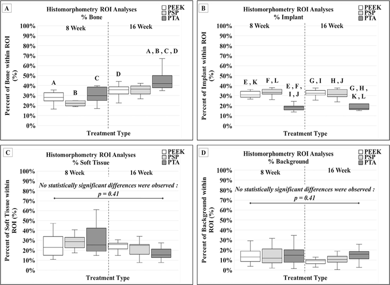
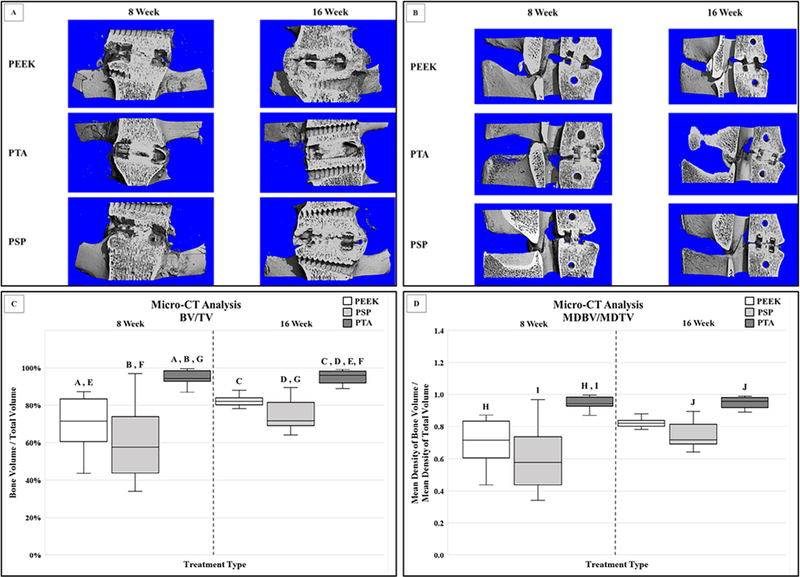
A skeletally mature ovine lumbar fusion model was used for this study. Interbody fusions were performed at L2-L3 and L4-L5 in 27 mature sheep using three different interbody cages (ie, polyetheretherketone [PEEK], plasma sprayed porous titanium-coated PEEK [PSP], and 3D-printed porous titanium alloy cage [PTA]). Non-destructive kinematic testing was performed in the three primary directions of motion. The specimens were then analyzed using micro-computed tomography (µ-CT); quantitative measures of the bony fusion were performed. Histomorphometric analyses were also performed in the sagittal plane through the interbody device. Outcome parameters were compared between cage designs and time points.
Flexion-extension range of motion (ROM) was statistically reduced for the PTA group compared with the PEEK cages at 16 weeks (p-value=.02). Only the PTA cages demonstrated a statistically significant decrease in ROM and increase in stiffness across all three loading directions between the 8-week and 16-week sacrifice time points (p-value≤.01). Micro-CT data demonstrated significantly greater total bone volume within the graft window for the PTA cages at both 8 weeks and 16 weeks compared with the PEEK cages (p-value<.01).
A direct comparison of interbody implants demonstrates significant and measurable differences in biomechanical, µ-CT, and histologic performance in an ovine model. The 3D-printed porous titanium interbody cage resulted in statistically significant reductions in ROM, increases in the bone ingrowth profile, as well as average construct stiffness compared with PEEK and PSP.
脊柱外科中使用的椎间融合器材料存在显著差异。理论上,使用多孔钛材料的 3D 打印椎间融合器可以提供更一致的骨长入和生物固定。
本研究旨在为脊柱融合用椎间材料的决策提供循证方法。
比较性动物研究。
本研究使用成熟的绵羊腰椎融合模型。在 27 只成熟绵羊的 L2-L3 和 L4-L5 处进行椎间融合,使用三种不同的椎间融合器(即聚醚醚酮[PEEK]、等离子喷涂多孔钛涂层 PEEK[PSP]和 3D 打印多孔钛合金 cage[PTA])。在三个主要运动方向进行非破坏性运动学测试。然后使用微计算机断层扫描(µ-CT)对标本进行分析;对骨融合进行定量测量。还在矢状面通过椎间器械进行组织形态计量学分析。比较各组之间的 cage 设计和时间点的结果参数。
与 PEEK cage 相比,PTA cage 在 16 周时屈伸活动范围(ROM)的统计学降低(p 值=.02)。仅 PTA cage 在 8 周和 16 周的牺牲时间点之间,在所有三个加载方向上均表现出 ROM 统计学显著降低和刚度增加(p 值≤.01)。微 CT 数据表明,在 8 周和 16 周时,与 PEEK cage 相比,PTA cage 中的移植物窗口内的总骨体积明显更大(p 值<.01)。
对椎间植入物的直接比较表明,在绵羊模型中,生物力学、µ-CT 和组织学性能存在显著且可测量的差异。与 PEEK 和 PSP 相比,3D 打印多孔钛椎间融合器在 ROM 显著降低、骨长入 profile 增加以及平均构建刚度方面均具有统计学意义。