• 文献检索
  • 文档翻译
  • 深度研究
  • 学术资讯
  • Suppr Zotero 插件Zotero 插件
  • 邀请有礼
  • 套餐&价格
  • 历史记录
应用&插件
Suppr Zotero 插件Zotero 插件浏览器插件Mac 客户端Windows 客户端微信小程序
定价
高级版会员购买积分包购买API积分包
服务
文献检索文档翻译深度研究API 文档MCP 服务
关于我们
关于 Suppr公司介绍联系我们用户协议隐私条款
关注我们

Suppr 超能文献

核心技术专利:CN118964589B侵权必究
粤ICP备2023148730 号-1Suppr @ 2026

文献检索

告别复杂PubMed语法,用中文像聊天一样搜索,搜遍4000万医学文献。AI智能推荐,让科研检索更轻松。

立即免费搜索

文件翻译

保留排版,准确专业,支持PDF/Word/PPT等文件格式,支持 12+语言互译。

免费翻译文档

深度研究

AI帮你快速写综述,25分钟生成高质量综述,智能提取关键信息,辅助科研写作。

立即免费体验

结合常规超声和对比增强超声得到的原发肿瘤特征有助于预测乳腺影像报告和数据系统 4 类恶性乳腺病变中腋窝淋巴结阳性。

Combining primary tumor features derived from conventional and contrast-enhanced ultrasound facilitates the prediction of positive axillary lymph nodes in Breast Imaging Reporting and Data System category 4 malignant breast lesions.

机构信息

Department of Ultrasound, The First Affiliated Hospital of Nanjing Medical University, Nanjing, China.

Department of Breast Surgery, The First Affiliated Hospital of Nanjing Medical University, Nanjing, China.

出版信息

Diagn Interv Radiol. 2023 May 31;29(3):469-477. doi: 10.4274/dir.2022.22534. Epub 2023 Jan 25.

DOI:10.4274/dir.2022.22534
PMID:36994900
原文链接:https://pmc.ncbi.nlm.nih.gov/articles/PMC10679605/
Abstract

PURPOSE

To determine whether the primary tumor features derived from conventional ultrasound (US) and contrast-enhanced US (CEUS) facilitate the prediction of positive axillary lymph nodes (ALNs) in breast cancer diagnosed as Breast Imaging Reporting and Data System (BI-RADS) category 4.

METHODS

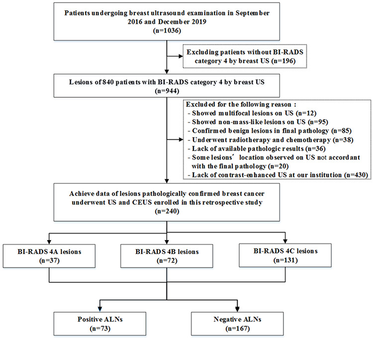
A total of 240 women with breast cancer who underwent preoperative conventional US, strain elastography, and CEUS between September 2016 and December 2019 were included. The multiple parameters of the primary tumor were obtained, and univariate and multivariate analyses were performed to predict positive ALNs. Then three prediction models (conventional US features, CEUS features, and the combined features) were developed, and the diagnostic performance was evaluated with receiver operating characteristic curves.

RESULTS

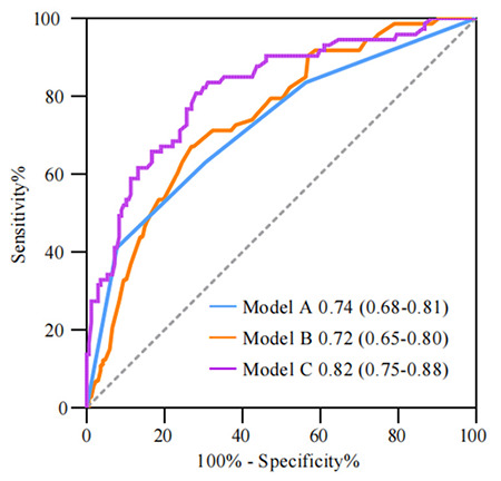
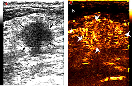
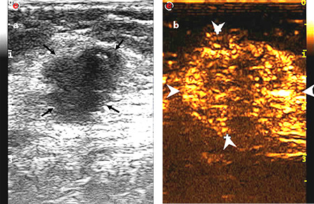
On conventional US, the traits of large size and the non-circumscribed margin of the primary tumor were marked as two independent predictors. On CEUS, the features of vessel perforation or distortion and the enhanced range of the primary tumor were marked as two independent predictors for positive ALNs. Three prediction models were then developed: model A (conventional US features), model B (CEUS features), and model C (model A plus B). Model C yielded the highest area under the curve (AUC) of 0.82 [95% confidence interval (CI), 0.75-0.88] compared with model A (AUC 0.74; 95% CI, 0.68-0.81; = 0.008) and model B (AUC 0.72; 95% CI, 0.65-0.80; < 0.001) as per the DeLong test.

CONCLUSION

CEUS, as a non-invasive examination technique, can be used to predict ALN metastasis. Combining conventional US and CEUS may produce favorable predictive accuracy for positive ALNs in BI-RADS category 4 breast cancer.

摘要

目的

确定源于常规超声(US)和对比增强超声(CEUS)的原发肿瘤特征是否有助于预测经乳腺影像报告和数据系统(BI-RADS)分类为 4 类的乳腺癌患者的阳性腋窝淋巴结(ALN)。

方法

共纳入 240 例于 2016 年 9 月至 2019 年 12 月期间接受术前常规 US、应变弹性成像和 CEUS 检查的乳腺癌患者。获得原发肿瘤的多个参数,并进行单因素和多因素分析以预测阳性 ALN。然后建立了三个预测模型(常规 US 特征、CEUS 特征和联合特征),并通过受试者工作特征曲线评估诊断性能。

结果

在常规 US 上,原发肿瘤的大尺寸和非边界清晰的特征被标记为两个独立的预测因素。在 CEUS 上,血管穿孔或扭曲和原发肿瘤强化范围的特征被标记为阳性 ALN 的两个独立预测因素。然后建立了三个预测模型:模型 A(常规 US 特征)、模型 B(CEUS 特征)和模型 C(模型 A 加 B)。模型 C 的曲线下面积(AUC)最高,为 0.82 [95%置信区间(CI),0.75-0.88],与模型 A(AUC 为 0.74;95%CI,0.68-0.81; = 0.008)和模型 B(AUC 为 0.72;95%CI,0.65-0.80; < 0.001)相比,根据 DeLong 检验有统计学差异。

结论

CEUS 作为一种非侵入性检查技术,可用于预测 ALN 转移。结合常规 US 和 CEUS 可能会提高 BI-RADS 4 类乳腺癌阳性 ALN 的预测准确性。

https://cdn.ncbi.nlm.nih.gov/pmc/blobs/437b/10679605/c8b43ddf7105/DIR-29-469-g6.jpg
https://cdn.ncbi.nlm.nih.gov/pmc/blobs/437b/10679605/7d1bf3656f84/DIR-29-469-g1.jpg
https://cdn.ncbi.nlm.nih.gov/pmc/blobs/437b/10679605/27c787909f0c/DIR-29-469-g2.jpg
https://cdn.ncbi.nlm.nih.gov/pmc/blobs/437b/10679605/de486f139c67/DIR-29-469-g3.jpg
https://cdn.ncbi.nlm.nih.gov/pmc/blobs/437b/10679605/ede6f6d911ab/DIR-29-469-g4.jpg
https://cdn.ncbi.nlm.nih.gov/pmc/blobs/437b/10679605/1296c262bf67/DIR-29-469-g5.jpg
https://cdn.ncbi.nlm.nih.gov/pmc/blobs/437b/10679605/c8b43ddf7105/DIR-29-469-g6.jpg

相似文献

1
Combining primary tumor features derived from conventional and contrast-enhanced ultrasound facilitates the prediction of positive axillary lymph nodes in Breast Imaging Reporting and Data System category 4 malignant breast lesions.结合常规超声和对比增强超声得到的原发肿瘤特征有助于预测乳腺影像报告和数据系统 4 类恶性乳腺病变中腋窝淋巴结阳性。
Diagn Interv Radiol. 2023 May 31;29(3):469-477. doi: 10.4274/dir.2022.22534. Epub 2023 Jan 25.
2
Adding contrast-enhanced ultrasound markers to conventional axillary ultrasound improves specificity for predicting axillary lymph node metastasis in patients with breast cancer.在常规腋窝超声中添加增强超声标志物可提高预测乳腺癌患者腋窝淋巴结转移的特异性。
Br J Radiol. 2021 Feb 1;94(1118):20200874. doi: 10.1259/bjr.20200874. Epub 2020 Dec 22.
3
Combining the Ultrasound Features of Primary Tumor and Axillary Lymph Nodes Can Reduce False-Negative Rate during the Prediction of High Axillary Node Burden in BI-RADS Category 4 or 5 Breast Cancer Lesions.联合原发性肿瘤和腋窝淋巴结的超声特征可降低 BI-RADS 4 或 5 类乳腺癌病变中预测腋窝淋巴结高负荷的假阴性率。
Ultrasound Med Biol. 2020 Aug;46(8):1941-1948. doi: 10.1016/j.ultrasmedbio.2020.04.003. Epub 2020 May 22.
4
Predictive value of contrast-enhanced ultrasonography and ultrasound elastography for management of BI-RADS category 4 nonpalpable breast masses.超声造影及超声弹性成像对 BI-RADS 分类 4 类触诊阴性乳腺肿块的预测价值。
Eur J Radiol. 2024 Apr;173:111391. doi: 10.1016/j.ejrad.2024.111391. Epub 2024 Feb 28.
5
Value of Contrast-Enhanced Ultrasound Combined with Immune-Inflammatory Markers in Predicting Axillary Lymph Node Metastasis of Breast Cancer.超声造影联合免疫炎症标志物对乳腺癌腋窝淋巴结转移的预测价值。
Acad Radiol. 2024 Sep;31(9):3535-3545. doi: 10.1016/j.acra.2024.06.013. Epub 2024 Jun 25.
6
Diagnosis of sub-centimetre breast lesions: combining BI-RADS-US with strain elastography and contrast-enhanced ultrasound-a preliminary study in China.乳腺亚厘米级病变的诊断:联合乳腺影像报告和数据系统-超声(BI-RADS-US)、应变弹性成像及超声造影在中国的初步研究
Eur Radiol. 2017 Jun;27(6):2443-2450. doi: 10.1007/s00330-016-4628-4. Epub 2016 Oct 19.
7
Application of contrast-enhanced ultrasound in the diagnosis of small breast lesions.超声造影在乳腺小病灶诊断中的应用
Clin Hemorheol Microcirc. 2018;70(3):291-300. doi: 10.3233/CH-170368.
8
Multimodal Ultrasound Imaging in Breast Imaging-Reporting and Data System 4 Breast Lesions: A Prediction Model for Malignancy.多模态超声成像在乳腺影像报告和数据系统 4 类乳腺病变中的应用:恶性肿瘤预测模型。
Ultrasound Med Biol. 2020 Dec;46(12):3188-3199. doi: 10.1016/j.ultrasmedbio.2020.08.003. Epub 2020 Sep 4.
9
A Prospective Multicenter Study on the Additive Value of Contrast-Enhanced Ultrasound for Biopsy Decision of Ultrasound BI-RADS 4 Breast Lesions.对比增强超声在超声 BI-RADS 4 类乳腺病变活检决策中附加价值的前瞻性多中心研究。
Ultrasound Med Biol. 2024 Aug;50(8):1224-1231. doi: 10.1016/j.ultrasmedbio.2024.04.010. Epub 2024 May 24.
10
The clinical value of conventional ultrasound combined with contrast-enhanced ultrasound in the evaluation of BI-RADS 4 lesions detected by magnetic resonance imaging.常规超声联合超声造影在磁共振成像检测 BI-RADS 4 病变评估中的临床价值。
Br J Radiol. 2022 Aug 1;95(1136):20220025. doi: 10.1259/bjr.20220025. Epub 2022 May 30.

引用本文的文献

1
Early Warning of Axillary Lymph Node Metastasis in Breast Cancer Patients Using Multi-Omics Signature: A Machine Learning-Based Retrospective Study.基于多组学特征的乳腺癌患者腋窝淋巴结转移早期预警:一项基于机器学习的回顾性研究
Int J Gen Med. 2024 Dec 12;17:6101-6114. doi: 10.2147/IJGM.S499238. eCollection 2024.
2
A nomogram based on conventional and contrast-enhanced ultrasound radiomics for the noninvasively prediction of axillary lymph node metastasis in breast cancer patients.一种基于常规超声和超声造影影像学特征的列线图,用于无创预测乳腺癌患者腋窝淋巴结转移。
Front Oncol. 2024 May 10;14:1400872. doi: 10.3389/fonc.2024.1400872. eCollection 2024.

本文引用的文献

1
Utility of Ultrasound Strain Elastography to Differentiate Benign from Malignant Lesions of the Breast.超声应变弹性成像在鉴别乳腺良恶性病变中的应用价值。
J Med Ultrasound. 2020 Sep 5;29(2):89-93. doi: 10.4103/JMU.JMU_32_20. eCollection 2021 Apr-Jun.
2
Adding contrast-enhanced ultrasound markers to conventional axillary ultrasound improves specificity for predicting axillary lymph node metastasis in patients with breast cancer.在常规腋窝超声中添加增强超声标志物可提高预测乳腺癌患者腋窝淋巴结转移的特异性。
Br J Radiol. 2021 Feb 1;94(1118):20200874. doi: 10.1259/bjr.20200874. Epub 2020 Dec 22.
3
Lymph Node Metastasis Prediction from Primary Breast Cancer US Images Using Deep Learning.
基于深度学习的原发性乳腺癌 US 图像淋巴结转移预测。
Radiology. 2020 Jan;294(1):19-28. doi: 10.1148/radiol.2019190372. Epub 2019 Nov 19.
4
Correlation Between Contrast-enhanced Ultrasound Characteristics (Qualitative and Quantitative) and Pathological Prognostic Factors in Breast Cancer.乳腺癌超声造影特征(定性和定量)与病理预后因素的相关性
In Vivo. 2018 Jul-Aug;32(4):945-954. doi: 10.21873/invivo.11333.
5
The relationship between tumour size, nodal status and distant metastases: on the origins of breast cancer.肿瘤大小、淋巴结状态和远处转移之间的关系:乳腺癌的起源。
Breast Cancer Res Treat. 2018 Aug;170(3):647-656. doi: 10.1007/s10549-018-4796-9. Epub 2018 Apr 24.
6
Meta-Analysis: Contrast-Enhanced Ultrasound Versus Conventional Ultrasound for Differentiation of Benign and Malignant Breast Lesions.荟萃分析:对比增强超声与传统超声在鉴别乳腺良恶性病变中的应用
Ultrasound Med Biol. 2018 May;44(5):919-929. doi: 10.1016/j.ultrasmedbio.2018.01.022. Epub 2018 Mar 9.
7
Correlation between Ultrasound Appearance of Small Breast Cancer and Axillary Lymph Node Metastasis.小乳腺癌的超声表现与腋窝淋巴结转移的相关性
Ultrasound Med Biol. 2018 Feb;44(2):342-349. doi: 10.1016/j.ultrasmedbio.2017.09.020. Epub 2017 Nov 14.
8
Role of BI-RADS Ultrasound Subcategories 4A to 4C in Predicting Breast Cancer.BI-RADS 超声分类 4A 至 4C 在预测乳腺癌中的作用。
Clin Breast Cancer. 2018 Aug;18(4):e507-e511. doi: 10.1016/j.clbc.2017.09.002. Epub 2017 Sep 19.
9
Association between US features of primary tumor and axillary lymph node metastasis in patients with clinical T1-T2N0 breast cancer.临床T1-T2N0期乳腺癌患者原发肿瘤超声特征与腋窝淋巴结转移的相关性
Acta Radiol. 2018 Apr;59(4):402-408. doi: 10.1177/0284185117723039. Epub 2017 Jul 27.
10
Regional Contrast-Enhanced Ultrasonography (CEUS) Characteristics of Breast Cancer and Correlation with Microvessel Density (MVD).乳腺癌的区域对比增强超声(CEUS)特征及其与微血管密度(MVD)的相关性
Med Sci Monit. 2017 Jul 14;23:3428-3436. doi: 10.12659/msm.901734.