Viral Immunopathology Laboratory, Infection, Immunity and Inflammation Research Theme, School of Medical Sciences, Faculty of Medicine and Health, The University of Sydney, Sydney, NSW, 2006, Australia.
Sydney Cytometry, The University of Sydney and Centenary Institute, Sydney, NSW, 2006, Australia.
Acta Neuropathol Commun. 2023 Apr 4;11(1):60. doi: 10.1186/s40478-023-01547-4.
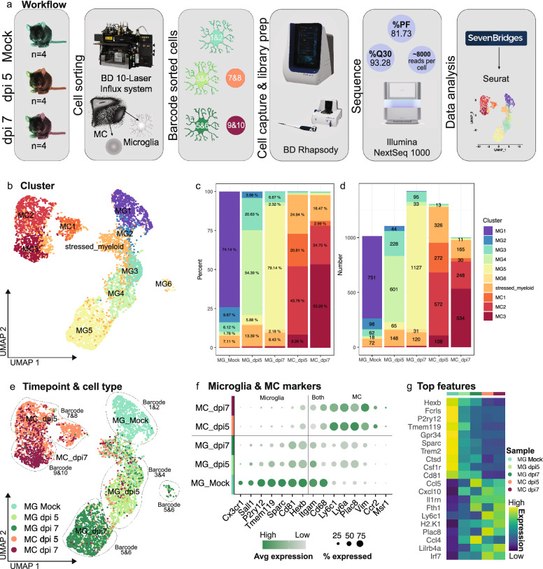
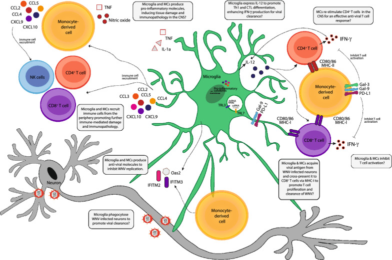
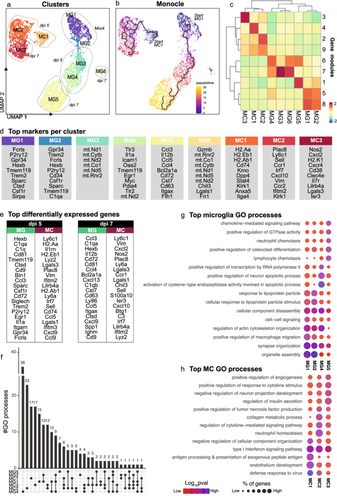
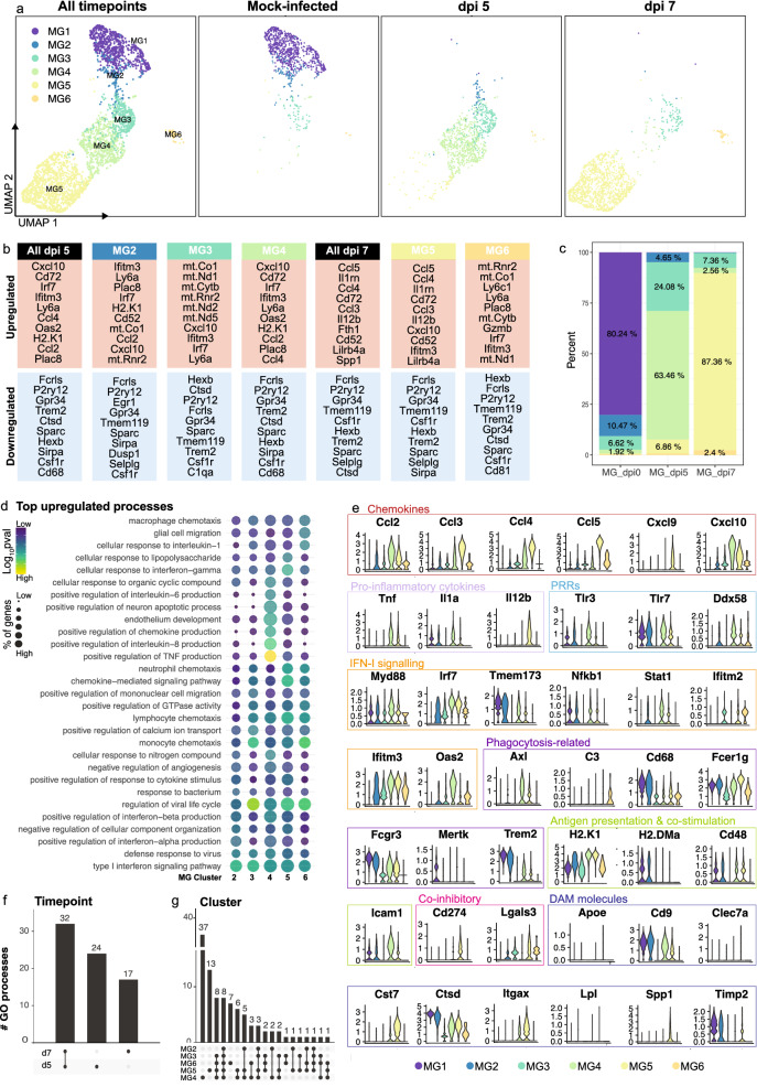
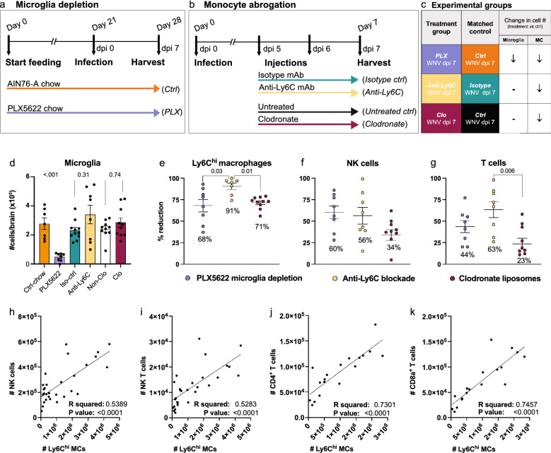
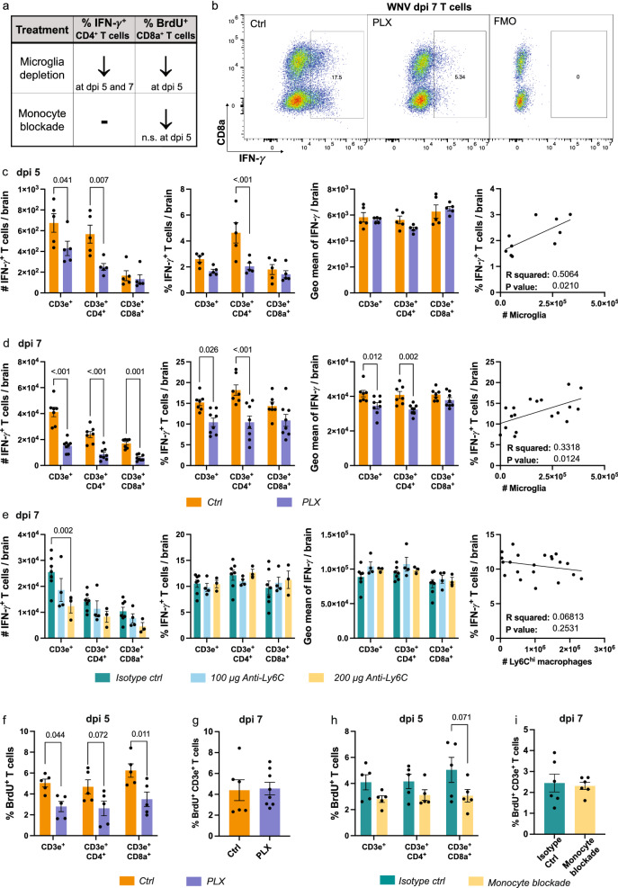
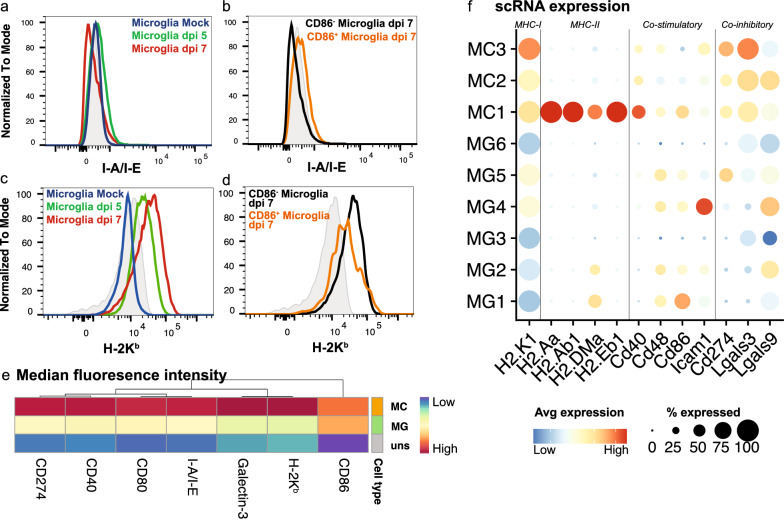
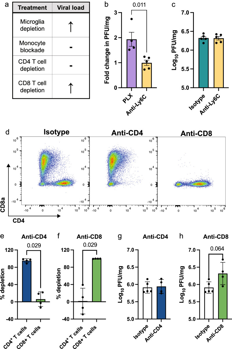
As the resident parenchymal myeloid population in the central nervous system (CNS), microglia are strategically positioned to respond to neurotropic virus invasion and have been implicated in promoting both disease resolution and progression in the acute and post-infectious phase of virus encephalitis. In a mouse model of West Nile virus encephalitis (WNE), infection of the CNS results in recruitment of large numbers of peripheral immune cells into the brain, the majority being nitric oxide (NO)-producing Ly6C inflammatory monocyte-derived cells (MCs). In this model, these cells enhance immunopathology and mortality. However, the contribution of microglia to this response is currently undefined. Here we used a combination of experimental tools, including single-cell RNA sequencing (scRNA-seq), microglia and MC depletion reagents, high-dimensional spectral cytometry and computational algorithms to dissect the differential contribution of microglia and MCs to the anti-viral immune response in severe neuroinflammation seen in WNE. Intriguingly, analysis of scRNA-seq data revealed 6 unique microglia and 3 unique MC clusters that were predominantly timepoint-specific, demonstrating substantial transcriptional adaptation with disease progression over the course of WNE. While microglia and MC adopted unique gene expression profiles, gene ontology enrichment analysis, coupled with microglia and MC depletion studies, demonstrated a role for both of these cells in the trafficking of peripheral immune cells into the CNS, T cell responses and viral clearance. Over the course of infection, microglia transitioned from a homeostatic to an anti-viral and then into an immune cell-recruiting phenotype. Conversely, MC adopted antigen-presenting, immune cell-recruiting and NO-producing phenotypes, which all had anti-viral function. Overall, this study defines for the first time the single-cell transcriptomic responses of microglia and MCs over the course of WNE, demonstrating both protective and pathological roles of these cells that could potentially be targeted for differential therapeutic intervention to dampen immune-mediated pathology, while maintaining viral clearance functions.
作为中枢神经系统(CNS)中常驻实质髓系细胞,小胶质细胞处于响应神经亲和性病毒入侵的战略位置,并被牵连到促进病毒脑炎的急性期和感染后阶段疾病的缓解和进展。在西尼罗河病毒脑炎(WNE)的小鼠模型中,CNS 的感染导致大量外周免疫细胞招募到大脑中,其中大多数是产生一氧化氮(NO)的 Ly6C 炎症性单核细胞衍生细胞(MC)。在该模型中,这些细胞增强了免疫病理学和死亡率。然而,小胶质细胞对该反应的贡献目前尚不清楚。在这里,我们使用了一系列实验工具,包括单细胞 RNA 测序(scRNA-seq)、小胶质细胞和 MC 耗竭试剂、高维光谱细胞术和计算算法,以剖析在 WNE 中所见的严重神经炎症中的抗病毒免疫反应中小胶质细胞和 MC 的差异贡献。有趣的是,scRNA-seq 数据分析揭示了 6 个独特的小胶质细胞和 3 个独特的 MC 簇,这些簇主要是时间点特异性的,表明随着 WNE 的发展,转录有了实质性的适应。虽然小胶质细胞和 MC 采用了独特的基因表达谱,但基因本体富集分析,加上小胶质细胞和 MC 耗竭研究,表明这两种细胞都在将外周免疫细胞运送到 CNS、T 细胞反应和病毒清除中发挥作用。在感染过程中,小胶质细胞从稳态转变为抗病毒状态,然后转变为招募免疫细胞的表型。相反,MC 采用了抗原呈递、免疫细胞募集和产生 NO 的表型,所有这些表型都具有抗病毒功能。总的来说,这项研究首次定义了 WNE 中小胶质细胞和 MC 的单细胞转录组反应,证明了这些细胞的保护性和病理性作用,这些作用可能成为靶向治疗干预的目标,以减轻免疫介导的病理学,同时保持病毒清除功能。