• 文献检索
  • 文档翻译
  • 深度研究
  • 学术资讯
  • Suppr Zotero 插件Zotero 插件
  • 邀请有礼
  • 套餐&价格
  • 历史记录
应用&插件
Suppr Zotero 插件Zotero 插件浏览器插件Mac 客户端Windows 客户端微信小程序
定价
高级版会员购买积分包购买API积分包
服务
文献检索文档翻译深度研究API 文档MCP 服务
关于我们
关于 Suppr公司介绍联系我们用户协议隐私条款
关注我们

Suppr 超能文献

核心技术专利:CN118964589B侵权必究
粤ICP备2023148730 号-1Suppr @ 2026

文献检索

告别复杂PubMed语法,用中文像聊天一样搜索,搜遍4000万医学文献。AI智能推荐,让科研检索更轻松。

立即免费搜索

文件翻译

保留排版,准确专业,支持PDF/Word/PPT等文件格式,支持 12+语言互译。

免费翻译文档

深度研究

AI帮你快速写综述,25分钟生成高质量综述,智能提取关键信息,辅助科研写作。

立即免费体验

脾脏硬化性血管瘤样结节性转化:多模态影像学特征及文献复习。

Sclerosing angiomatoid nodular transformation of the spleen: multimodality imaging features and literature review.

机构信息

Department of Radiology, The Second Affiliated Hospital of Fujian Medical University, No. 34 Zhongshan Bei Road, Licheng District, Quanzhou, Fujian, China.

Department of Radiology, Zhongshan Hospital, Fudan University, No. 180 Fenglin Road, Xuhui District, Shanghai, 200032, China.

出版信息

BMC Med Imaging. 2023 Apr 6;23(1):50. doi: 10.1186/s12880-023-01008-3.

DOI:10.1186/s12880-023-01008-3
PMID:37024862
原文链接:https://pmc.ncbi.nlm.nih.gov/articles/PMC10080891/
Abstract

OBJECTIVE

The purpose of this study was to evaluate the CT and MRI findings, clinicopathologic features, and differential diagnosis of Sclerosing angiomatoid nodular transformation (SANT).

METHODS AND MATERIALS

Seven men and seven women with pathological diagnoses of SANT were included in this retrospect study. Patients underwent at least one radiological examination before surgery. The number, shape, margin, size, attenuation, signal intensity, homogeneity, and enhancing pattern of the lesion were evaluated by two abdominal radiologists independently. Immunohistochemistry reports were available for 11 patients. The immunoreactivity to the vascular markers CD8, CD31, and CD34 was assessed.

RESULTS

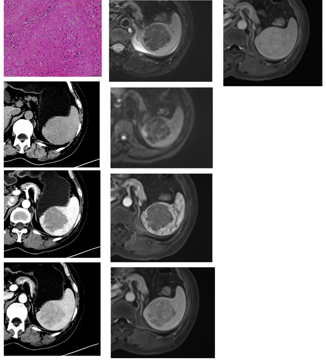
The 14 SANT patients (7 men, 7 women; mean age, 43.5 years; age range, 24-56 years) presented with a single lesion and showed no specific clinical symptoms. Among 14 patients, 12 patients underwent MR scan, 5 patients underwent CT scan and 3 patients underwent PET-CT. On CT, all 5 lesions showed hypodensity on non-contrast images and spoke-wheel enhancing pattern after contrast administration, and calcification was observed. On T2WI, 10 cases(83.3%)showed hypointensity and 2 cases (16.7%) showed hyperintensity with central hypointensity. On T1WI, 10 cases (83.3%) were isointense and 2 cases (16.7%) were slightly hypointense. 10 cases (83.3%) showed hypointensity on DWI and 2 cases (16.7%) showed slightly hyperintensity on DWI. After contrast administration, all 12 lesions showed progressive enhancement. 18 F-fluorodeoxyglucose (FDG) uptake in the tumor was seen in all three cases that underwent PET-CT. The maximum standardized uptake value (SUVmax) was 4.5, 5.1, and 3.8 respectively.

RESULTS

Apart from the progressive spoke-wheel enhancing pattern, DWI and ADC findings will add value to the diagnosis of SANT.

摘要

目的

本研究旨在评估硬化性血管样结节转化(SANT)的 CT 和 MRI 表现、临床病理特征及鉴别诊断。

方法和材料

本回顾性研究纳入了 7 名男性和 7 名女性病理诊断为 SANT 的患者。患者在术前均至少进行了 1 次影像学检查。由 2 名腹部放射科医生独立评估病变的数量、形状、边界、大小、衰减、信号强度、均匀性和增强模式。11 例患者的免疫组化报告可用,评估了其对血管标志物 CD8、CD31 和 CD34 的免疫反应性。

结果

14 例 SANT 患者(7 名男性,7 名女性;平均年龄 43.5 岁;年龄范围 24-56 岁)表现为单个病灶,无特异性临床症状。14 例患者中,12 例行 MR 扫描,5 例行 CT 扫描,3 例行 PET-CT 扫描。CT 上,所有 5 个病灶在非增强图像上呈低信号,增强后呈辐轮样强化,且可见钙化。T2WI 上,10 例(83.3%)呈低信号,2 例(16.7%)呈中心低信号的高信号。T1WI 上,10 例(83.3%)为等信号,2 例(16.7%)为稍低信号。10 例(83.3%)DWI 呈低信号,2 例(16.7%)DWI 呈稍高信号。增强后,所有 12 个病灶均呈渐进性强化。3 例行 PET-CT 检查的患者均可见肿瘤 18F-氟脱氧葡萄糖(FDG)摄取,最大标准化摄取值(SUVmax)分别为 4.5、5.1 和 3.8。

结果

除渐进性辐轮样强化模式外,DWI 和 ADC 表现将有助于 SANT 的诊断。

https://cdn.ncbi.nlm.nih.gov/pmc/blobs/d616/10080891/1183c6d7643e/12880_2023_1008_Fig2_HTML.jpg
https://cdn.ncbi.nlm.nih.gov/pmc/blobs/d616/10080891/c1d152549f02/12880_2023_1008_Fig1_HTML.jpg
https://cdn.ncbi.nlm.nih.gov/pmc/blobs/d616/10080891/1183c6d7643e/12880_2023_1008_Fig2_HTML.jpg
https://cdn.ncbi.nlm.nih.gov/pmc/blobs/d616/10080891/c1d152549f02/12880_2023_1008_Fig1_HTML.jpg
https://cdn.ncbi.nlm.nih.gov/pmc/blobs/d616/10080891/1183c6d7643e/12880_2023_1008_Fig2_HTML.jpg

相似文献

1
Sclerosing angiomatoid nodular transformation of the spleen: multimodality imaging features and literature review.脾脏硬化性血管瘤样结节性转化:多模态影像学特征及文献复习。
BMC Med Imaging. 2023 Apr 6;23(1):50. doi: 10.1186/s12880-023-01008-3.
2
CT and MRI features of sclerosing angiomatoid nodular transformation of the spleen: A report of 18 patients with pathologic correlation.脾脏硬化性血管瘤样结节性转化的CT与MRI特征:18例有病理对照的病例报告
Diagn Interv Imaging. 2021 Jun;102(6):389-396. doi: 10.1016/j.diii.2021.01.003. Epub 2021 Jan 22.
3
Sclerosing Angiomatoid Nodular Transformation of the Spleen: Radiological Findings and Radiological-pathological Correlation.脾脏硬化性血管样结节性转化:放射学表现与放射病理学相关性。
Curr Med Imaging. 2024;20:e260423216201. doi: 10.2174/1573405620666230426122828.
4
Imaging Features of Sclerosing Angiomatoid Nodular Transformation in Spleen.脾脏硬化性血管瘤样结节性转化的影像学特征
J Comput Assist Tomogr. 2019 Nov/Dec;43(6):863-869. doi: 10.1097/RCT.0000000000000910.
5
An unusual case of sclerosing angiomatoid nodular transformation: radiological and histopathological analyses.一例不典型性血管肌脂瘤样结节性转化:影像学和组织病理学分析。
Turk J Med Sci. 2014;44(3):530-3.
6
Sclerosing angiomatoid nodular transformation (SANT) of the spleen: a case report with FDG-PET findings and literature review.脾脏硬化性血管瘤样结节性转化(SANT):一例伴有FDG-PET检查结果的病例报告及文献综述
Acta Radiol Open. 2016 Aug 10;5(8):2058460116649799. doi: 10.1177/2058460116649799. eCollection 2016 Aug.
7
18F-FDG avid Sclerosing Angiomatoid Nodular Transformation (SANT) of spleen on PET-CT - a rare mimicker of metastasis.PET-CT上脾脏的18F-FDG摄取阳性硬化性血管瘤样结节性转化(SANT)——一种罕见的转移瘤模拟病变。
Nucl Med Rev Cent East Eur. 2018;21(1):53. doi: 10.5603/NMR.2018.0014.
8
Sclerosing Angiomatoid Nodular Transformation of the Spleen: A Case Report.脾脏硬化性血管样结节性转化:一例报告。
Tokai J Exp Clin Med. 2020 Dec 20;45(4):236-242.
9
Sclerosing angiomatoid nodular transformation of the spleen: CT and MRI features with pathologic correlation.脾脏硬化性血管样结节性转化:与病理相关性的 CT 和 MRI 特征。
AJR Am J Roentgenol. 2013 Apr;200(4):W353-60. doi: 10.2214/AJR.12.9522.
10
CT and MRI findings of sclerosing angiomatoid nodular transformation of the spleen: spoke wheel pattern.脾脏硬化性血管瘤样结节性转化的CT和MRI表现:辐条轮模式。
Korean J Radiol. 2008 Jul;9 Suppl(Suppl):S52-5. doi: 10.3348/kjr.2008.9.s.s52.

引用本文的文献

1
Case Report: Sclerosing angiomatoid nodular transformation of the spleen: surgery or conservative treatment?病例报告:脾脏硬化性血管瘤样结节性转化:手术治疗还是保守治疗?
Front Immunol. 2025 Jul 28;16:1530363. doi: 10.3389/fimmu.2025.1530363. eCollection 2025.
2
Sclerosing angiomatoid nodular transformation of the spleen: clinical, computed tomography, and magnetic resonance imaging characteristics.脾脏硬化性血管瘤样结节性转化:临床、计算机断层扫描及磁共振成像特征
Quant Imaging Med Surg. 2025 Mar 3;15(3):1888-1897. doi: 10.21037/qims-24-1660. Epub 2025 Feb 26.
3
A case of sclerosing angiomatoid nodular transformation with central calcification.

本文引用的文献

1
Sclerosing Angiomatoid Nodular Transformation of the Spleen: A Diagnostic Conundrum.脾脏硬化性血管瘤样结节性转化:一个诊断难题。
J Belg Soc Radiol. 2022 Apr 5;106(1):12. doi: 10.5334/jbsr.2689. eCollection 2022.
2
Diffusion-Weighted Imaging in Oncology: An Update.肿瘤学中的扩散加权成像:最新进展
Cancers (Basel). 2020 Jun 8;12(6):1493. doi: 10.3390/cancers12061493.
3
Imaging Features of Sclerosing Angiomatoid Nodular Transformation in Spleen.脾脏硬化性血管瘤样结节性转化的影像学特征
一例伴有中央钙化的硬化性血管瘤样结节性转化病例。
Radiol Case Rep. 2024 Dec 17;20(3):1380-1385. doi: 10.1016/j.radcr.2024.11.071. eCollection 2025 Mar.
4
The Spectrum of Solitary Benign Splenic Lesions-Imaging Clues for a Noninvasive Diagnosis.孤立性脾脏良性病变的谱——无创诊断的影像学线索
Diagnostics (Basel). 2023 Jun 20;13(12):2120. doi: 10.3390/diagnostics13122120.
J Comput Assist Tomogr. 2019 Nov/Dec;43(6):863-869. doi: 10.1097/RCT.0000000000000910.
4
Sclerosing angiomatoid nodular transformation (SANT) of the spleen: A case report on CT and MRI.脾脏硬化性血管瘤样结节性转化(SANT):一例CT和MRI病例报告
BJR Case Rep. 2019 Jan 31;5(2):20180036. doi: 10.1259/bjrcr.20180036. eCollection 2019 Jun.
5
Sclerosing angiomatoid nodular transformation in the spleen: A case series study and literature review.脾脏硬化性血管瘤样结节性转化:病例系列研究及文献综述
Medicine (Baltimore). 2019 Apr;98(17):e15154. doi: 10.1097/MD.0000000000015154.
6
Sclerosing Angiomatoid Nodular Transformation of the Spleen: Lessons from a Rare Case and Review of the Literature.脾脏硬化性血管瘤样结节性转化:来自一例罕见病例的经验及文献复习
Intern Med. 2019 May 15;58(10):1433-1441. doi: 10.2169/internalmedicine.1948-18. Epub 2019 Jan 10.
7
Sclerosing angiomatoid nodular transformation of the spleen mimicking metastasis of melanoma: a case report and review of the literature.脾脏硬化性血管瘤样结节性转化酷似黑色素瘤转移:1例报告并文献复习
J Med Case Rep. 2017 Sep 3;11(1):251. doi: 10.1186/s13256-017-1400-6.
8
Sclerosing angiomatoid nodular transformation (SANT) of the spleen: a case report with FDG-PET findings and literature review.脾脏硬化性血管瘤样结节性转化(SANT):一例伴有FDG-PET检查结果的病例报告及文献综述
Acta Radiol Open. 2016 Aug 10;5(8):2058460116649799. doi: 10.1177/2058460116649799. eCollection 2016 Aug.
9
The value of contrast-enhanced dynamic and diffusion-weighted MR imaging for distinguishing benign and malignant splenic masses.对比增强动态磁共振成像和扩散加权磁共振成像在鉴别脾脏良恶性肿块中的价值。
Br J Radiol. 2016 Jul;89(1063):20160054. doi: 10.1259/bjr.20160054. Epub 2016 May 10.
10
Two cases of sclerosing angiomatoid nodular transformation of the spleen with gradual growth: usefulness of diffusion-weighted imaging.两例脾脏硬化性血管瘤样结节性转化伴逐渐生长:扩散加权成像的应用价值
Clin Imaging. 2015 Mar-Apr;39(2):315-7. doi: 10.1016/j.clinimag.2014.10.015. Epub 2014 Oct 22.