Department of Radiology, Athinoula A. Martinos Center for Biomedical Imaging, Massachusetts General Hospital, Charlestown, Massachusetts, USA.
Department of Neuropathology, Massachusetts General Hospital, Boston, Massachusetts, USA.
Brain Pathol. 2023 Jul;33(4):e13159. doi: 10.1111/bpa.13159. Epub 2023 Apr 10.
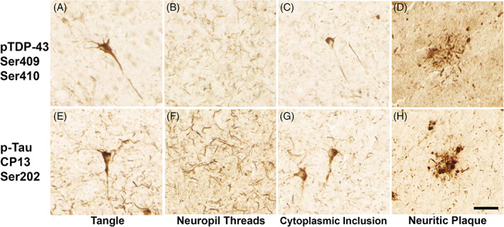
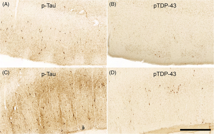
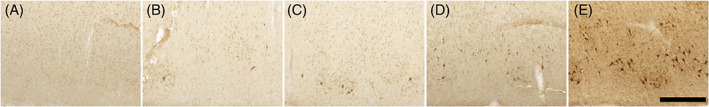
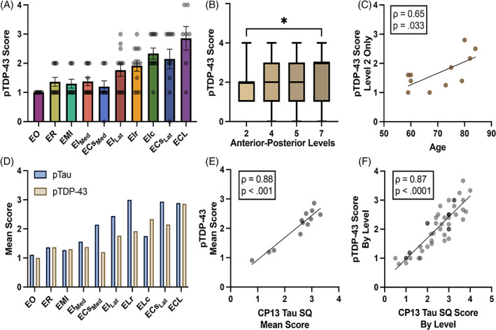
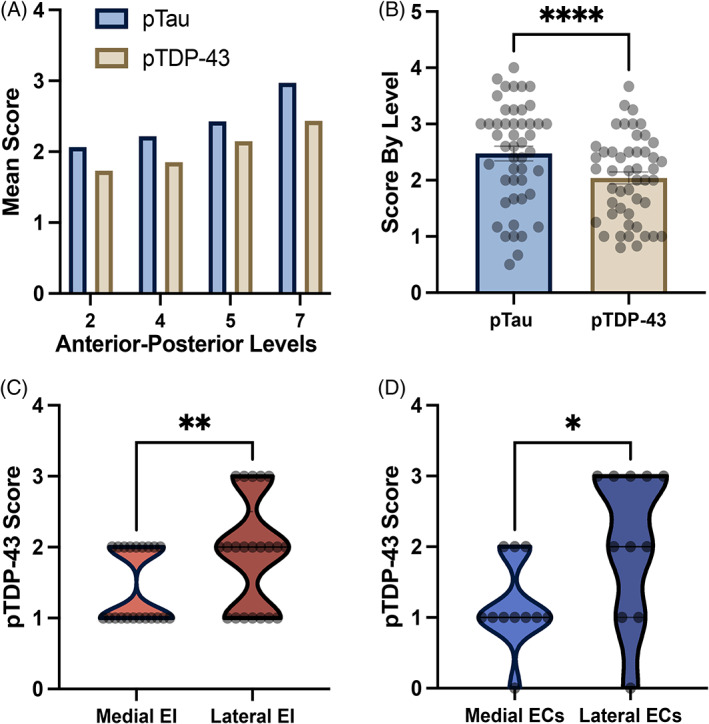
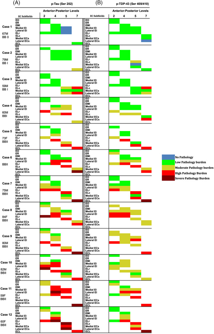
Phosphorylated tau (p-tau) pathology correlates strongly with cognitive decline and is a pathological hallmark of Alzheimer's Disease (AD). In recent years, phosphorylated transactive response DNA-binding protein (pTDP-43) has emerged as a common comorbidity, found in up to 70% of all AD cases (Josephs et al., Acta Neuropathol, 131(4), 571-585; Josephs, Whitwell, et al., Acta Neuropathol, 127(6), 811-824). Current staging schemes for pTDP-43 in AD and primary age-related tauopathy (PART) track its progression throughout the brain, but the distribution of pTDP-43 within the entorhinal cortex (EC) at the earliest stages has not been studied. Moreover, the exact nature of p-tau and pTDP-43 co-localization is debated. We investigated the selective vulnerability of the entorhinal subfields to phosphorylated pTDP-43 pathology in preclinical AD and PART postmortem tissue. Within the EC, posterior-lateral subfields showed the highest semi-quantitative pTDP-43 density scores, while the anterior-medial subfields had the lowest. On the rostrocaudal axis, pTDP-43 scores were higher posteriorly than anteriorly (p < 0.010), peaking at the posterior-most level (p < 0.050). Further, we showed the relationship between pTDP-43 and p-tau in these regions at pathology-positive but clinically silent stages. P-tau and pTDP-43 presented a similar pattern of affected subregions (p < 0.0001) but differed in density magnitude (p < 0.0001). P-tau burden was consistently higher than pTDP-43 at every anterior-posterior level and in most EC subfields. These findings highlight pTDP-43 burden heterogeneity within the EC and the posterior-lateral subfields as the most vulnerable regions within stage II of the current pTDP-43 staging schemes for AD and PART. The EC is a point of convergence for p-tau and pTDP-43 and identifying its most vulnerable neuronal populations will prove key for early diagnosis and disease intervention.
磷酸化 tau (p-tau) 病理学与认知能力下降密切相关,是阿尔茨海默病 (AD) 的病理学标志。近年来,磷酸化反式激活反应 DNA 结合蛋白 (pTDP-43) 已成为一种常见的合并症,在多达 70%的 AD 病例中发现(Josephs 等人,《神经病理学杂志》,131(4),571-585;Josephs,Whitwell 等人,《神经病理学杂志》,127(6),811-824)。AD 和原发性年龄相关性 tau 病 (PART) 中 pTDP-43 的当前分期方案可跟踪其在大脑中的进展,但在最早阶段,pTDP-43 在内嗅皮层 (EC) 中的分布尚未研究。此外,p-tau 和 pTDP-43 共定位的确切性质存在争议。我们研究了在临床前 AD 和 PART 尸检组织中 pTDP-43 病理学对内嗅皮层亚区的选择性易感性。在内嗅皮层,后外侧亚区显示出最高的半定量 pTDP-43 密度评分,而前内侧亚区最低。在前后轴上,pTDP-43 评分后部高于前部(p<0.010),在后部达到峰值(p<0.050)。此外,我们在病理学阳性但临床无症状的阶段显示了这些区域中 pTDP-43 与 p-tau 之间的关系。p-tau 和 pTDP-43 表现出受影响亚区相似的模式(p<0.0001),但密度大小不同(p<0.0001)。p-tau 负担始终高于每个前后水平和大多数 EC 亚区的 pTDP-43。这些发现突出了 EC 内 pTDP-43 负担的异质性以及当前 AD 和 PART 的 pTDP-43 分期方案 II 中最脆弱的后外侧亚区。EC 是 p-tau 和 pTDP-43 的汇聚点,确定其最脆弱的神经元群体将是早期诊断和疾病干预的关键。