Casamitjana Adrià, Mancini Matteo, Robinson Eleanor, Peter Loïc, Annunziata Roberto, Althonayan Juri, Crampsie Shauna, Blackburn Emily, Billot Benjamin, Atzeni Alessia, Puonti Oula, Balbastre Yaël, Schmidt Peter, Hughes James, Augustinack Jean C, Edlow Brian L, Zöllei Lilla, Thomas David L, Kliemann Dorit, Bocchetta Martina, Strand Catherine, Holton Janice L, Jaunmuktane Zane, Iglesias Juan Eugenio
Department of Medical Physics and Biomedical Engineering, University College London, London, United Kingdom.
Research Institute of Computer Vision and Robotics, University of Girona, Girona, Spain.
bioRxiv. 2024 Sep 6:2024.02.05.579016. doi: 10.1101/2024.02.05.579016.
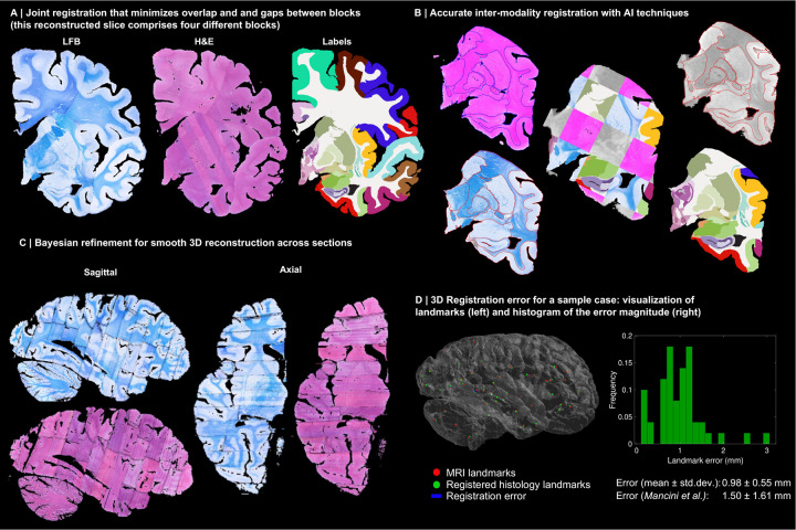
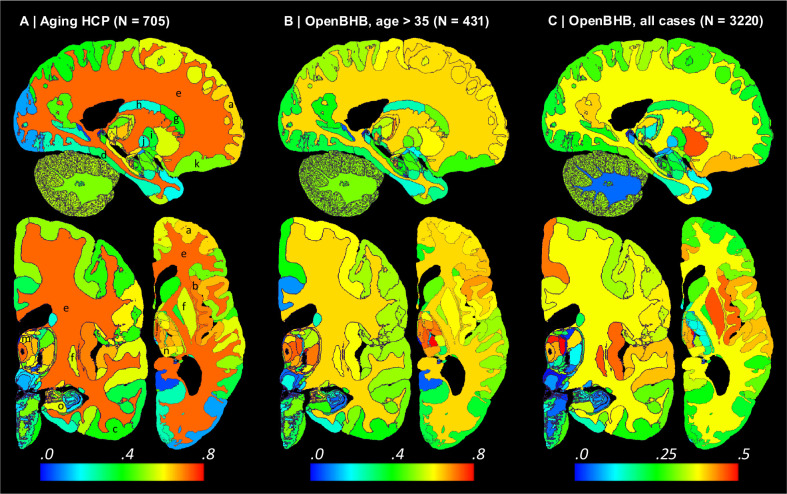
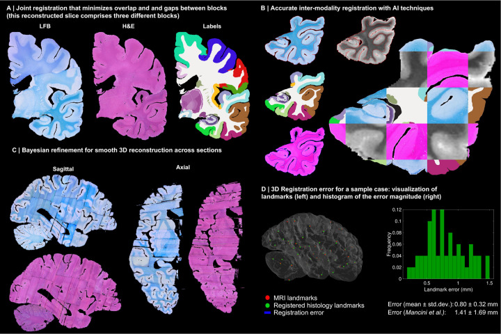
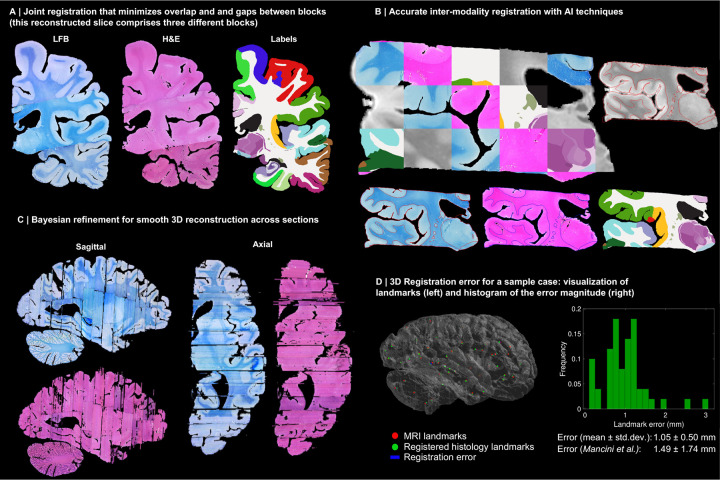
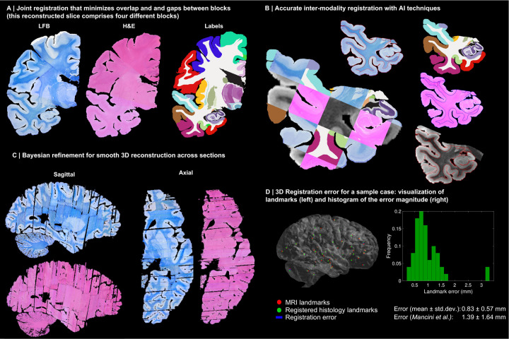
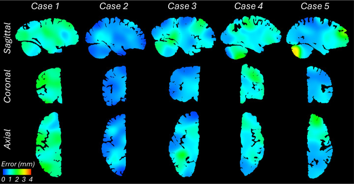
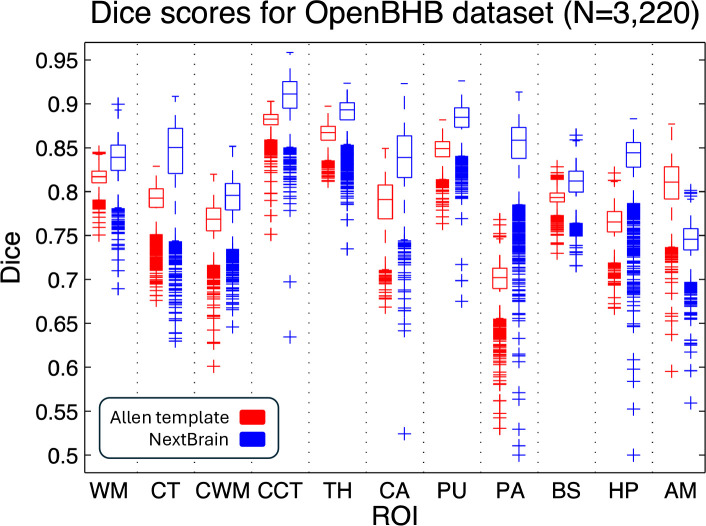
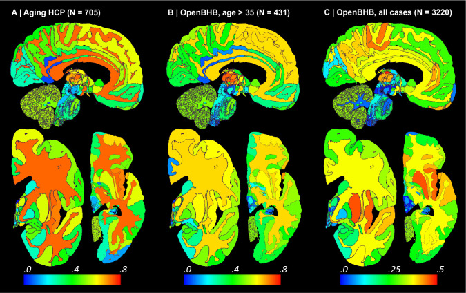
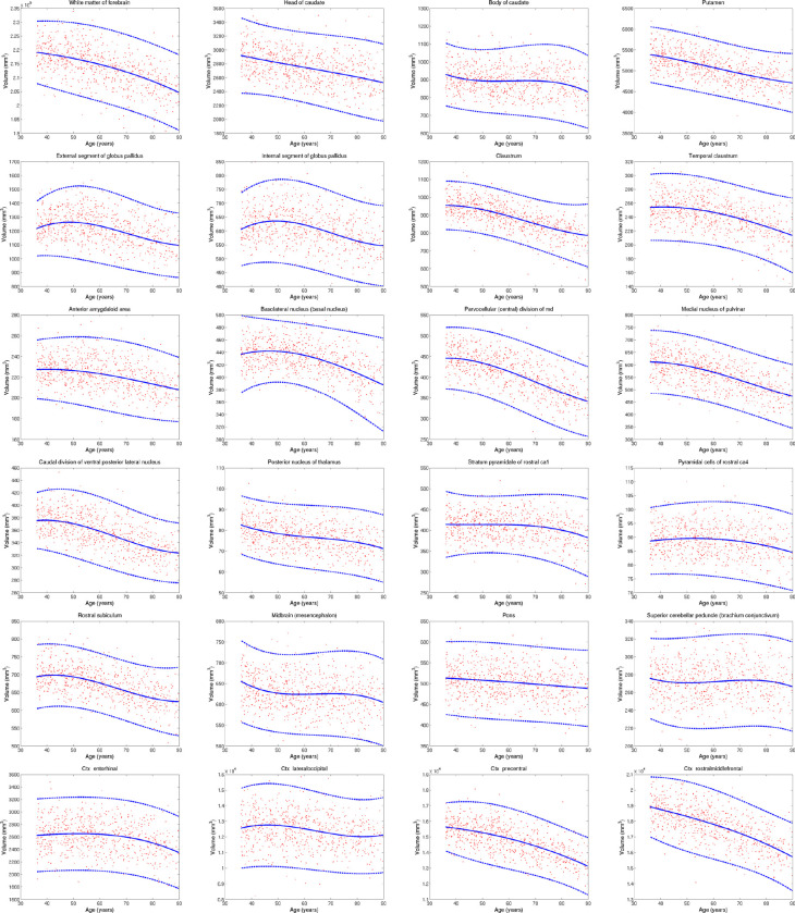
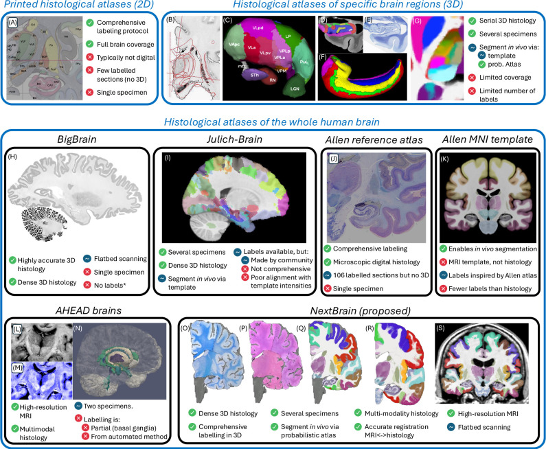
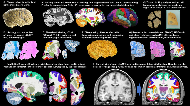
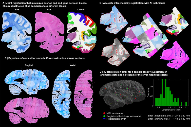
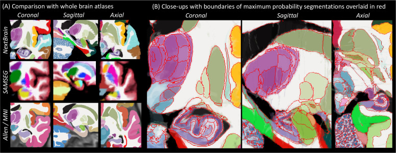
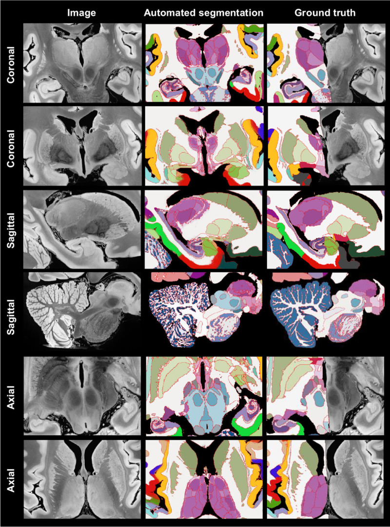
Magnetic resonance imaging (MRI) is the standard tool to image the human brain In this domain, digital brain atlases are essential for subject-specific segmentation of anatomical regions of interest (ROIs) and spatial comparison of neuroanatomy from different subjects in a common coordinate frame. High-resolution, digital atlases derived from histology (e.g., Allen atlas [7], BigBrain [13], Julich [15]), are currently the state of the art and provide exquisite 3D cytoarchitectural maps, but lack probabilistic labels throughout the whole brain. Here we present a next-generation probabilistic atlas of human brain anatomy built from serial 3D histology and corresponding highly granular delineations of five whole brain hemispheres. We developed AI techniques to align and reconstruct ~10,000 histological sections into coherent 3D volumes with joint geometric constraints (no overlap or gaps between sections), as well as to semi-automatically trace the boundaries of 333 distinct anatomical ROIs on all these sections. Comprehensive delineation on multiple cases enabled us to build Further, we created a companion Bayesian tool for automated segmentation of the 333 ROIs in any or brain MRI scan using the atlas. We showcase two applications of the atlas: automated segmentation of ultra-high-resolution MRI and volumetric analysis of Alzheimer's disease and healthy brain ageing based on ~4,000 publicly available MRI scans. We publicly release: the raw and aligned data (including an online visualisation tool); the probabilistic atlas; the segmentation tool; and ground truth delineations for a 100 μm isotropic hemisphere (that we use for quantitative evaluation of our segmentation method in this paper). By enabling researchers worldwide to analyse brain MRI scans at a superior level of granularity without manual effort or highly specific neuroanatomical knowledge, holds promise to increase the specificity of MRI findings and ultimately accelerate our quest to understand the human brain in health and disease.
磁共振成像(MRI)是对人类大脑进行成像的标准工具。在这一领域,数字脑图谱对于感兴趣的解剖区域(ROI)的特定主体分割以及在共同坐标框架下不同主体神经解剖结构的空间比较至关重要。源自组织学的高分辨率数字图谱(例如艾伦图谱[7]、大脑图谱[13]、尤利希图谱[15])是目前的先进水平,提供了精美的三维细胞结构图谱,但缺乏全脑的概率性标签。在此,我们展示了一个基于连续三维组织学和五个全脑半球的相应高分辨率描绘构建的人类脑解剖结构的下一代概率图谱。我们开发了人工智能技术,将约10,000个组织学切片对齐并重建为具有联合几何约束(切片之间无重叠或间隙)的连贯三维体积,并半自动追踪所有这些切片上333个不同解剖ROI的边界。对多个病例的全面描绘使我们能够构建……此外,我们创建了一个配套的贝叶斯工具,用于使用该图谱对任何大脑MRI扫描中的333个ROI进行自动分割。我们展示了该图谱的两个应用:超高分辨率MRI的自动分割以及基于约4000份公开可用MRI扫描对阿尔茨海默病和健康脑老化的体积分析。我们公开发布:原始数据和对齐后的数据(包括在线可视化工具);概率图谱;分割工具;以及一个100μm各向同性半球的真实边界描绘(我们在本文中用于对分割方法进行定量评估)。通过使全球研究人员能够在无需人工努力或高度特定的神经解剖学知识的情况下以更高的粒度水平分析大脑MRI扫描,该图谱有望提高MRI结果的特异性,并最终加速我们对健康和疾病状态下人类大脑的理解探索。