State Key Laboratory of Ophthalmology, Zhongshan Ophthalmic Center, Sun Yat-Sen University, Guangdong Provincial Key Laboratory of Ophthalmology and Visual Science, Guangzhou, China.
Invest Ophthalmol Vis Sci. 2023 Apr 3;64(4):12. doi: 10.1167/iovs.64.4.12.
The proliferation, migration, and epithelial-mesenchymal transition (EMT) of lens epithelial cells (LECs) are believed to be the pathological mechanisms underlying anterior subcapsular cataract (ASC). Bone morphogenetic proteins (BMPs) inhibit transforming growth factor-beta (TGF-β)-induced fibrosis in the lens. Herein, we aimed to further clarify the roles of BMP-4/BMP-7 in the progression and the underlying mechanisms of fibrotic cataract.
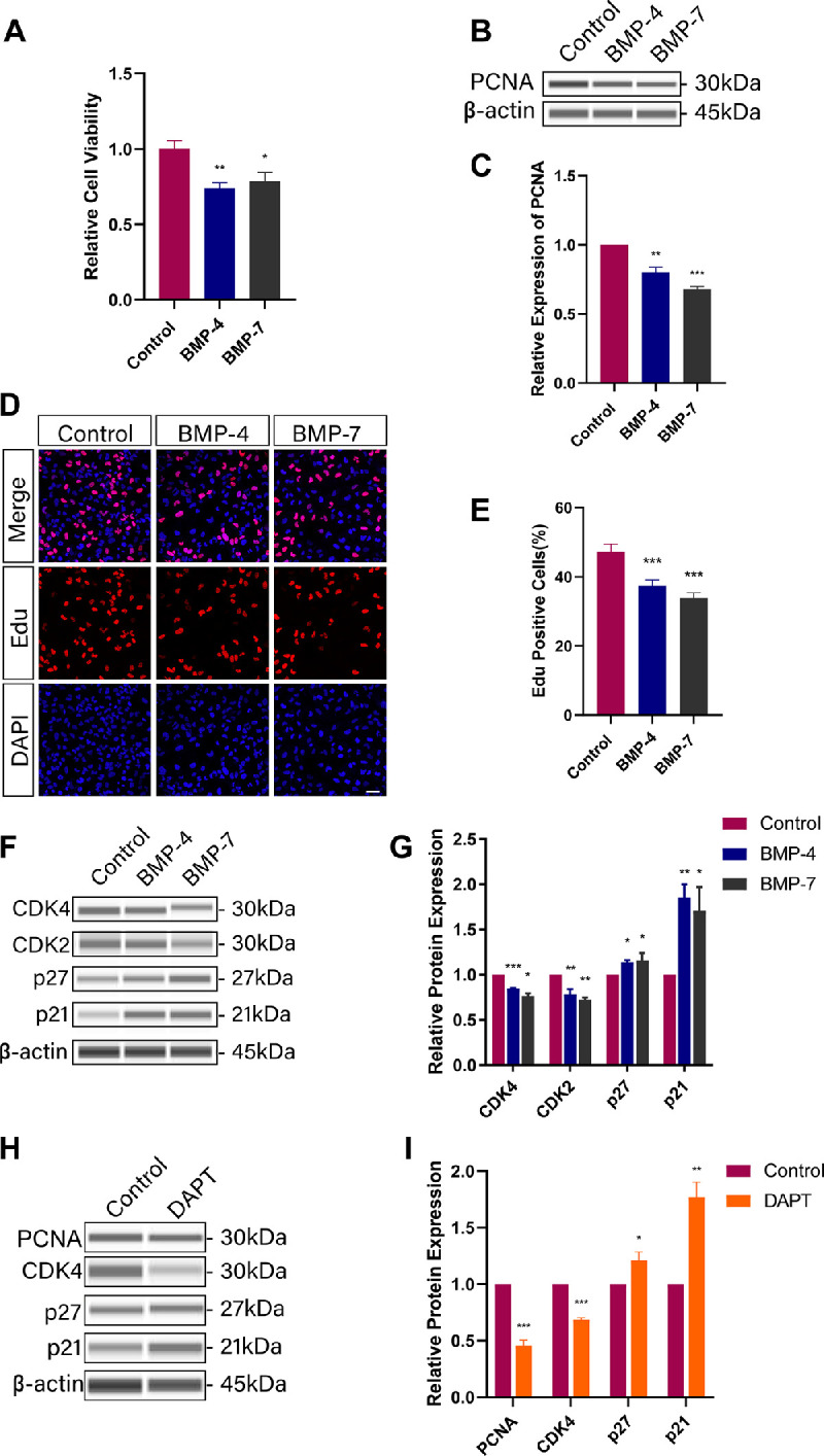
BMP-4/BMP-7, TGF-β2, jagged-1 peptide, or DAPT were applied in a mouse injury-induced ASC model and in the human LEC cell line SRA01/04. The volume of opacity was examined by a slit lamp and determined by lens anterior capsule whole-mount immunofluorescence. Global gene expression changes were assessed by RNA sequencing, and the levels of individual mRNAs were validated by real-time PCR. Protein expression was determined by the Simple Western sample dilution buffer. Cell proliferation was examined by CCK8 and EdU assays, and cell migration was measured by Transwell and wound healing assays.
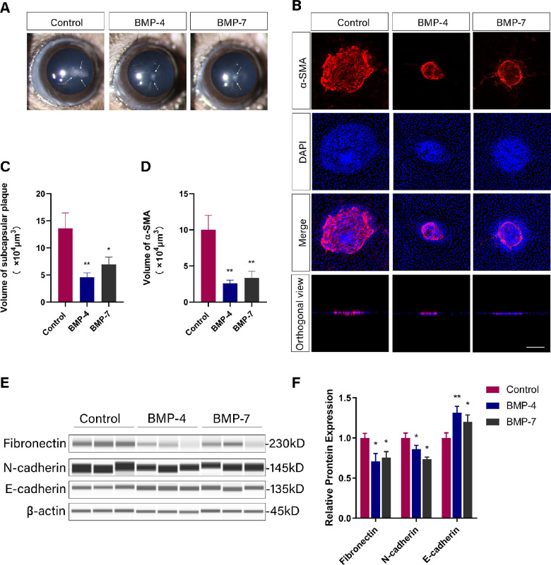
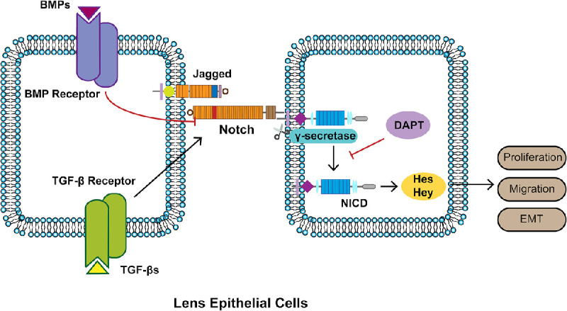
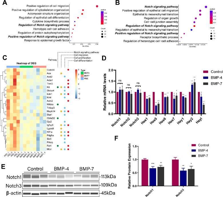
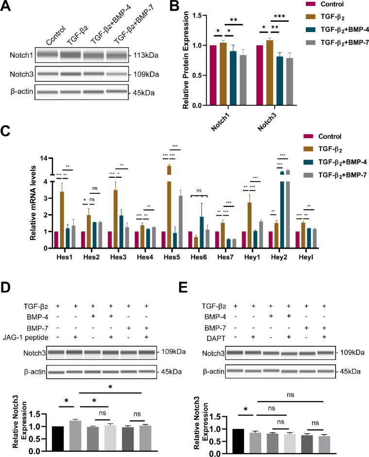
Anterior chamber injection of BMP-4/BMP-7 significantly suppressed subcapsular opacification formation. RNA sequencing of the mouse ASC model identified the Notch pathway as a potential mechanism involved in BMP-mediated inhibition of ASC. Consistently, BMP-4/BMP-7 selectively suppressed Notch1 and Notch3 and their downstream genes, including Hes and Hey. BMP-4/BMP-7 or DAPT suppressed cell proliferation by inducing G1 cell cycle arrest. BMP-4/BMP-7 also inhibited TGF-β2-induced cell migration and EMT by modulating the Notch pathway.
BMP-4/BMP-7 attenuated ASC by inhibiting proliferation, migration, and EMT of LECs via modulation of the Notch pathway, thereby providing a new avenue for ASC treatment.
晶状体上皮细胞(LEC)的增殖、迁移和上皮-间充质转化(EMT)被认为是前囊下白内障(ASC)的病理机制。骨形态发生蛋白(BMPs)抑制晶状体中转化生长因子-β(TGF-β)诱导的纤维化。在此,我们旨在进一步阐明 BMP-4/BMP-7 在纤维性白内障进展中的作用及其潜在机制。
在小鼠损伤诱导的 ASC 模型和人 LEC 细胞系 SRA01/04 中应用 BMP-4/BMP-7、TGF-β2、Jagged-1 肽或 DAPT。用裂隙灯检查混浊程度,并通过晶状体前囊全层免疫荧光法确定。通过 RNA 测序评估整体基因表达变化,并通过实时 PCR 验证个别 mRNA 的水平。用 Simple Western 样品稀释缓冲液测定蛋白质表达。通过 CCK8 和 EdU 测定法检测细胞增殖,通过 Transwell 和划痕愈合测定法测量细胞迁移。
前房注射 BMP-4/BMP-7 可显著抑制囊下混浊形成。对小鼠 ASC 模型的 RNA 测序鉴定出 Notch 通路是 BMP 介导的 ASC 抑制的潜在机制。一致地,BMP-4/BMP-7 选择性地抑制 Notch1 和 Notch3 及其下游基因,包括 Hes 和 Hey。BMP-4/BMP-7 或 DAPT 通过诱导 G1 细胞周期阻滞抑制细胞增殖。BMP-4/BMP-7 还通过调节 Notch 通路抑制 TGF-β2 诱导的细胞迁移和 EMT。
BMP-4/BMP-7 通过调节 Notch 通路抑制 LEC 的增殖、迁移和 EMT,从而减轻 ASC,为 ASC 的治疗提供了新途径。