• 文献检索
  • 文档翻译
  • 深度研究
  • 学术资讯
  • Suppr Zotero 插件Zotero 插件
  • 邀请有礼
  • 套餐&价格
  • 历史记录
应用&插件
Suppr Zotero 插件Zotero 插件浏览器插件Mac 客户端Windows 客户端微信小程序
定价
高级版会员购买积分包购买API积分包
服务
文献检索文档翻译深度研究API 文档MCP 服务
关于我们
关于 Suppr公司介绍联系我们用户协议隐私条款
关注我们

Suppr 超能文献

核心技术专利:CN118964589B侵权必究
粤ICP备2023148730 号-1Suppr @ 2026

文献检索

告别复杂PubMed语法,用中文像聊天一样搜索,搜遍4000万医学文献。AI智能推荐,让科研检索更轻松。

立即免费搜索

文件翻译

保留排版,准确专业,支持PDF/Word/PPT等文件格式,支持 12+语言互译。

免费翻译文档

深度研究

AI帮你快速写综述,25分钟生成高质量综述,智能提取关键信息,辅助科研写作。

立即免费体验

体外至体内外推和高内涵成像技术同时用于化学诱导肝脂肪变性和肝毒性标志物的特征描述。

In vitro to in vivo extrapolation and high-content imaging for simultaneous characterization of chemically induced liver steatosis and markers of hepatotoxicity.

机构信息

Department of Health Sciences and Technology, ETH Zurich, Schmelzbergstrasse 9, 8092, Zurich, Switzerland.

School of Life Sciences, University of Applied Sciences and Arts Northwestern Switzerland, 4132, Muttenz, Switzerland.

出版信息

Arch Toxicol. 2023 Jun;97(6):1701-1721. doi: 10.1007/s00204-023-03490-8. Epub 2023 Apr 12.

DOI:10.1007/s00204-023-03490-8
PMID:37046073
原文链接:https://pmc.ncbi.nlm.nih.gov/articles/PMC10182956/
Abstract

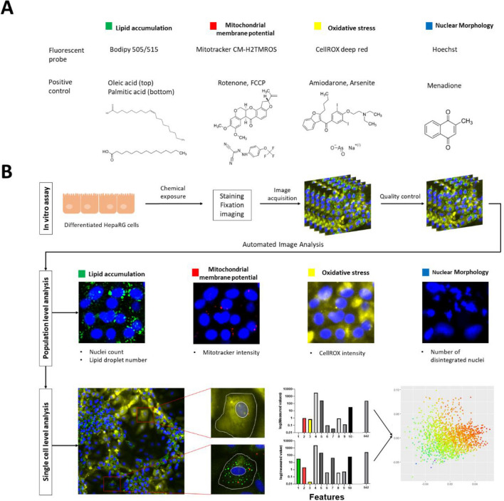
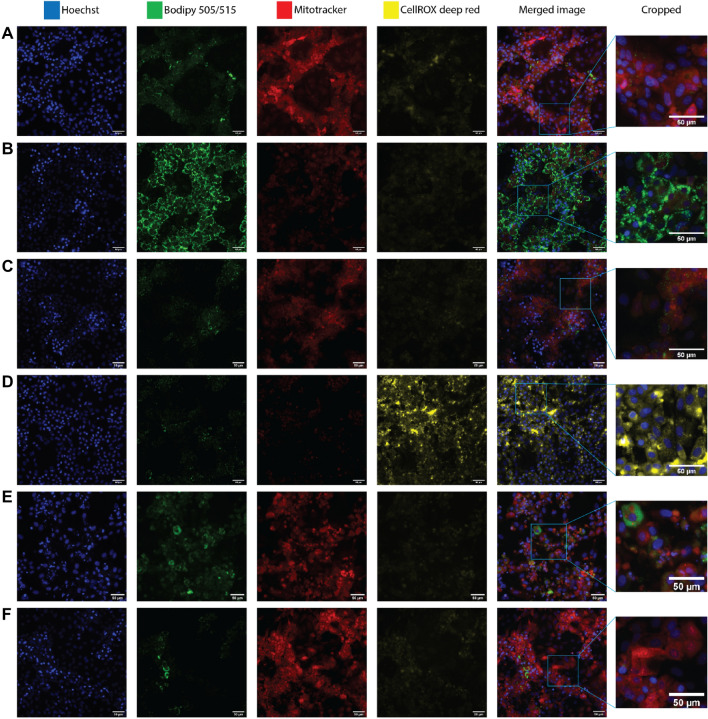
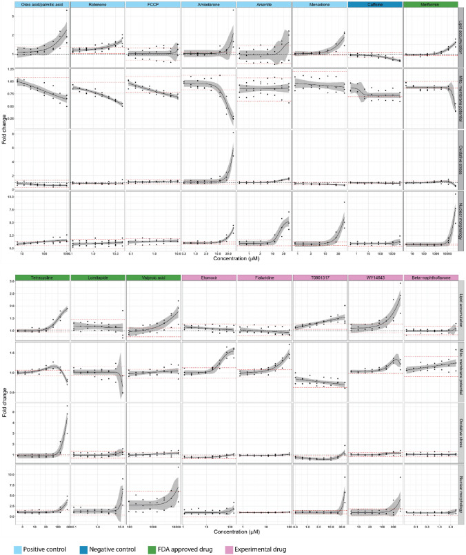
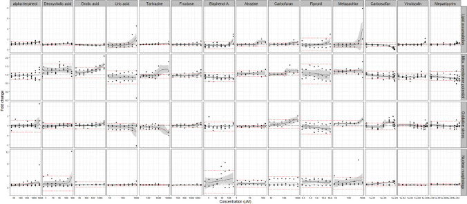
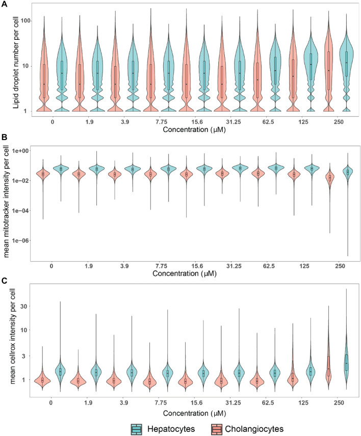
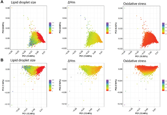
Chemically induced steatosis is characterized by lipid accumulation associated with mitochondrial dysfunction, oxidative stress and nucleus distortion. New approach methods integrating in vitro and in silico models are needed to identify chemicals that may induce these cellular events as potential risk factors for steatosis and associated hepatotoxicity. In this study we used high-content imaging for the simultaneous quantification of four cellular markers as sentinels for hepatotoxicity and steatosis in chemically exposed human liver cells in vitro. Furthermore, we evaluated the results with a computational model for the extrapolation of human oral equivalent doses (OED). First, we tested 16 reference chemicals with known capacities to induce cellular alterations in nuclear morphology, lipid accumulation, mitochondrial membrane potential and oxidative stress. Then, using physiologically based pharmacokinetic modeling and reverse dosimetry, OEDs were extrapolated from data of any stimulated individual sentinel response. The extrapolated OEDs were confirmed to be within biologically relevant exposure ranges for the reference chemicals. Next, we tested 14 chemicals found in food, selected from thousands of putative chemicals on the basis of structure-based prediction for nuclear receptor activation. Amongst these, orotic acid had an extrapolated OED overlapping with realistic exposure ranges. Thus, we were able to characterize known steatosis-inducing chemicals as well as data-scarce food-related chemicals, amongst which we confirmed orotic acid to induce hepatotoxicity. This strategy addresses needs of next generation risk assessment and can be used as a first chemical prioritization hazard screening step in a tiered approach to identify chemical risk factors for steatosis and hepatotoxicity-associated events.

摘要

化学诱导性脂肪变性的特征是脂质积累与线粒体功能障碍、氧化应激和核变形有关。需要整合体外和计算模型的新方法来识别可能诱导这些细胞事件的化学物质,这些细胞事件可能是脂肪变性和相关肝毒性的潜在风险因素。在这项研究中,我们使用高内涵成像技术同时定量检测四种细胞标志物,作为体外化学暴露人肝细胞肝毒性和脂肪变性的标志物。此外,我们还使用计算模型评估了这些结果,以推断人体口服等效剂量(OED)。首先,我们测试了 16 种已知具有改变核形态、脂质积累、线粒体膜电位和氧化应激能力的参考化学物质。然后,使用基于生理的药代动力学建模和反向剂量测定,从任何刺激的个体标志物反应数据推断 OED。推断的 OED 被证实处于参考化学物质的生物相关暴露范围内。接下来,我们测试了 14 种在食品中发现的化学物质,这些化学物质是基于核受体激活的结构预测从数千种潜在化学物质中选择的。在这些化学物质中,乳清酸的推断 OED 与现实暴露范围重叠。因此,我们能够对已知的脂肪变性诱导化学物质以及数据稀缺的食品相关化学物质进行特征描述,其中我们证实乳清酸会引起肝毒性。该策略满足下一代风险评估的需求,可作为一种分层方法中的第一步化学优先危害筛选步骤,以识别脂肪变性和相关肝毒性事件的化学风险因素。

https://cdn.ncbi.nlm.nih.gov/pmc/blobs/211f/10182956/345dfb956b1c/204_2023_3490_Fig8_HTML.jpg
https://cdn.ncbi.nlm.nih.gov/pmc/blobs/211f/10182956/4444307cc191/204_2023_3490_Fig1_HTML.jpg
https://cdn.ncbi.nlm.nih.gov/pmc/blobs/211f/10182956/a61727d56510/204_2023_3490_Fig2_HTML.jpg
https://cdn.ncbi.nlm.nih.gov/pmc/blobs/211f/10182956/f476bb7f1b00/204_2023_3490_Fig3_HTML.jpg
https://cdn.ncbi.nlm.nih.gov/pmc/blobs/211f/10182956/ff8590ec8e22/204_2023_3490_Fig4_HTML.jpg
https://cdn.ncbi.nlm.nih.gov/pmc/blobs/211f/10182956/a9be770fea8e/204_2023_3490_Fig5_HTML.jpg
https://cdn.ncbi.nlm.nih.gov/pmc/blobs/211f/10182956/21797aebcf61/204_2023_3490_Fig6_HTML.jpg
https://cdn.ncbi.nlm.nih.gov/pmc/blobs/211f/10182956/6218bf3d7db3/204_2023_3490_Fig7_HTML.jpg
https://cdn.ncbi.nlm.nih.gov/pmc/blobs/211f/10182956/345dfb956b1c/204_2023_3490_Fig8_HTML.jpg

相似文献

1
In vitro to in vivo extrapolation and high-content imaging for simultaneous characterization of chemically induced liver steatosis and markers of hepatotoxicity.体外至体内外推和高内涵成像技术同时用于化学诱导肝脂肪变性和肝毒性标志物的特征描述。
Arch Toxicol. 2023 Jun;97(6):1701-1721. doi: 10.1007/s00204-023-03490-8. Epub 2023 Apr 12.
2
In vitro/in silico prediction of drug induced steatosis in relation to oral doses and blood concentrations by the Nile Red assay.尼罗红法预测与口服剂量和血药浓度相关的药物诱导脂肪变性的体外/计算机研究。
Toxicol Lett. 2022 Sep 1;368:33-46. doi: 10.1016/j.toxlet.2022.08.006. Epub 2022 Aug 10.
3
Evaluation of the Potential Risk of Drugs to Induce Hepatotoxicity in Human-Relationships between Hepatic Steatosis Observed in Non-Clinical Toxicity Study and Hepatotoxicity in Humans.评估药物在人体中诱发肝毒性的潜在风险——非临床毒性研究中观察到的肝脂肪变性与人类肝毒性之间的关系。
Int J Mol Sci. 2017 Apr 12;18(4):810. doi: 10.3390/ijms18040810.
4
Busulfan induces steatosis in HepaRG cells but not in primary human hepatocytes: Possible explanations and implication for the prediction of drug-induced liver injury.白消安可诱导 HepaRG 细胞发生脂肪变性,但不会诱导原代人肝细胞发生脂肪变性:可能的解释及其对药物性肝损伤预测的影响。
Fundam Clin Pharmacol. 2024 Feb;38(1):152-167. doi: 10.1111/fcp.12951. Epub 2023 Sep 4.
5
Metabolomics-based strategy to assess drug hepatotoxicity and uncover the mechanisms of hepatotoxicity involved.基于代谢组学的策略来评估药物肝毒性,并揭示涉及的肝毒性机制。
Arch Toxicol. 2023 Jun;97(6):1723-1738. doi: 10.1007/s00204-023-03474-8. Epub 2023 Apr 6.
6
A biologically based model to quantitatively assess the role of the nuclear receptors liver X (LXR), and pregnane X (PXR) on chemically induced hepatic steatosis.建立一种基于生物学的模型,定量评估核受体肝 X(LXR)和妊娠相关 X 受体(PXR)在化学诱导性肝脂肪变性中的作用。
Toxicol Lett. 2022 Apr 15;359:46-54. doi: 10.1016/j.toxlet.2022.02.002. Epub 2022 Feb 7.
7
Advantageous use of HepaRG cells for the screening and mechanistic study of drug-induced steatosis.HepaRG细胞在药物性脂肪变性的筛选和机制研究中的优势应用。
Toxicol Appl Pharmacol. 2016 Jul 1;302:1-9. doi: 10.1016/j.taap.2016.04.007. Epub 2016 Apr 16.
8
Mechanisms of amiodarone and valproic acid induced liver steatosis in mouse in vivo act as a template for other hepatotoxicity models.胺碘酮和丙戊酸在小鼠体内诱导肝脂肪变性的机制可作为其他肝毒性模型的模板。
Arch Toxicol. 2014 Aug;88(8):1573-88. doi: 10.1007/s00204-014-1211-0. Epub 2014 Feb 18.
9
Estimating provisional margins of exposure for data-poor chemicals using high-throughput computational methods.使用高通量计算方法估算数据匮乏化学品的临时暴露限值。
Front Pharmacol. 2022 Oct 7;13:980747. doi: 10.3389/fphar.2022.980747. eCollection 2022.
10
QSAR Modeling of ToxCast Assays Relevant to the Molecular Initiating Events of AOPs Leading to Hepatic Steatosis.QSAR 模型构建——预测导致脂肪性肝病的 AOPs 的分子起始事件的 ToxCast 检测结果。
J Chem Inf Model. 2018 Aug 27;58(8):1501-1517. doi: 10.1021/acs.jcim.8b00297. Epub 2018 Jul 26.

引用本文的文献

1
A simple, reliable and easily generalizable cell-based assay for screening potential drugs that inhibit lipid accumulation.一种用于筛选抑制脂质积累的潜在药物的简单、可靠且易于推广的基于细胞的检测方法。
Curr Res Toxicol. 2024 Dec 26;8:100213. doi: 10.1016/j.crtox.2024.100213. eCollection 2025.
2
Understanding and using Animal Models of Hepatotoxicity.理解和使用肝毒性动物模型。
Curr Pharm Des. 2025;31(12):943-956. doi: 10.2174/0113816128338726241029175250.
3
Seminar: Functional Exposomics and Mechanisms of Toxicity-Insights from Model Systems and NAMs.

本文引用的文献

1
Estimating Hepatotoxic Doses Using High-Content Imaging in Primary Hepatocytes.使用原代肝细胞的高内涵成像技术估算肝毒性剂量。
Toxicol Sci. 2021 Sep 28;183(2):285-301. doi: 10.1093/toxsci/kfab091.
2
More than additive effects on liver triglyceride accumulation by combinations of steatotic and non-steatotic pesticides in HepaRG cells.在 HepaRG 细胞中,脂肪变性和非脂肪变性农药的组合对肝甘油三酯积累的影响大于相加作用。
Arch Toxicol. 2021 Apr;95(4):1397-1411. doi: 10.1007/s00204-021-02997-2. Epub 2021 Feb 11.
3
The implication of the air quality pattern in South Korea after the COVID-19 outbreak.
研讨会:功能外显组学与毒性作用机制——来自模型系统和新型替代方法的见解。
Environ Health Perspect. 2024 Sep;132(9):94201. doi: 10.1289/EHP13120. Epub 2024 Sep 4.
4
High-Resolution Mass Spectrometry for Human Exposomics: Expanding Chemical Space Coverage.高分辨率质谱技术在人类暴露组学中的应用:拓展化学空间覆盖范围。
Environ Sci Technol. 2024 Jul 23;58(29):12784-12822. doi: 10.1021/acs.est.4c01156. Epub 2024 Jul 10.
韩国 COVID-19 疫情爆发后空气质量模式的含义。
Sci Rep. 2020 Dec 31;10(1):22462. doi: 10.1038/s41598-020-80429-4.
4
O-GlcNAcylation: the "stress and nutrition receptor" in cell stress response.O-GlcNAc 修饰:细胞应激反应中的“应激和营养感受器”。
Cell Stress Chaperones. 2021 Mar;26(2):297-309. doi: 10.1007/s12192-020-01177-y. Epub 2020 Nov 7.
5
Drug-induced hepatic steatosis in absence of severe mitochondrial dysfunction in HepaRG cells: proof of multiple mechanism-based toxicity.在 HepaRG 细胞中无严重线粒体功能障碍的情况下药物诱导的肝脂肪变性:多种基于机制的毒性的证据。
Cell Biol Toxicol. 2021 Apr;37(2):151-175. doi: 10.1007/s10565-020-09537-1. Epub 2020 Jun 14.
6
Comparison of long-term versus short-term effects of okadaic acid on the apoptotic status of human HepaRG cells.比较冈田酸对人 HepaRG 细胞凋亡状态的长期和短期影响。
Chem Biol Interact. 2020 Feb 1;317:108937. doi: 10.1016/j.cbi.2020.108937. Epub 2020 Jan 8.
7
Perspectives on to Extrapolations.关于外推法的观点。 (你提供的原文“Perspectives on to Extrapolations.”表述似乎不太完整准确,我按照字面意思尽量贴近翻译了,你可以检查下原文是否准确。)
Appl In Vitro Toxicol. 2018 Dec 1;4(4):305-316. doi: 10.1089/aivt.2016.0026. Epub 2018 Dec 8.
8
Metformin reduces lipid accumulation in HepG2 cells via downregulation of miR-33b.二甲双胍通过下调 miR-33b 减少 HepG2 细胞中的脂质积累。
Arch Physiol Biochem. 2022 Apr;128(2):333-340. doi: 10.1080/13813455.2019.1680700. Epub 2019 Nov 5.
9
Lipid Droplets and the Management of Cellular Stress.脂滴与细胞应激管理。
Yale J Biol Med. 2019 Sep 20;92(3):435-452. eCollection 2019 Sep.
10
Augmentation of Glucotoxicity, Oxidative Stress, Apoptosis and Mitochondrial Dysfunction in HepG2 Cells by Palmitic Acid.软脂酸对 HepG2 细胞糖毒性、氧化应激、细胞凋亡及线粒体功能障碍的影响
Nutrients. 2019 Aug 22;11(9):1979. doi: 10.3390/nu11091979.